Muscular pains in the hip and knee
Understand common reasons for pain around the hip and knee, and how assessment, therapy and exercises can guide recovery.
Why do hip and knee hurt?
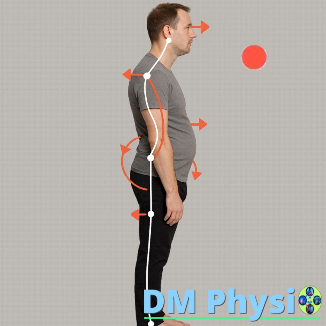
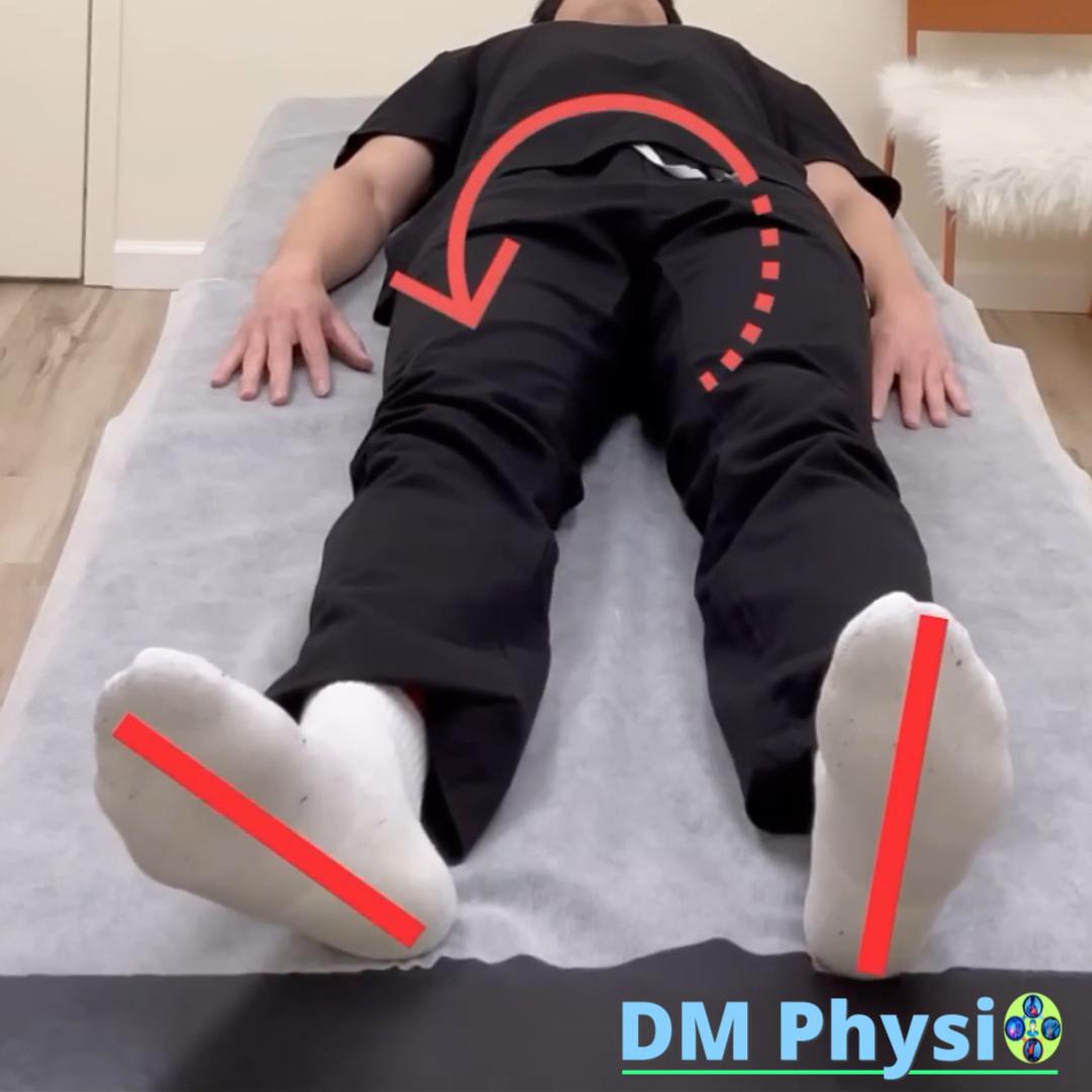
The pain most often comes from prolonged inappropriate positions - sitting, standing, walking or even sleeping in an uncomfortable position. Some muscles shorten, others stretch and weaken, which changes the gait and the way you step. This mis-distributes weight, irritates the tendons and tissues around the knee and hip, and causes pain – especially when squatting, climbing/descending stairs, getting up from a chair, or standing on one leg for long periods of time.
Muscle pain in the hip and knee is often confused with a "joint problem" (eg meniscus or osteoarthritis), but in many cases the cause is muscle and fascial dysfunction. Accurate functional assessment, habit correction, and targeted exercise are the keys to lasting relief.
How is muscle imbalance formed?
Improper posture
Sitting/standing for a long time or sleeping in an uncomfortable position "turns off" some muscles, and others become overworked and tight.
Compensation
When walking and climbing, the leg becomes "stiff" - the knee does not bend freely and the load is unevenly distributed.
Muscle spasm and pain
There are "nodules" (trigger points), limitation of movement, clicking and pain around the knee and in the thigh.
Which muscles are most commonly affected?
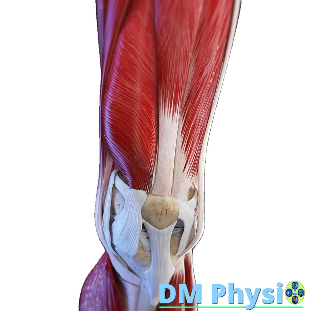
m. quadriceps femoris
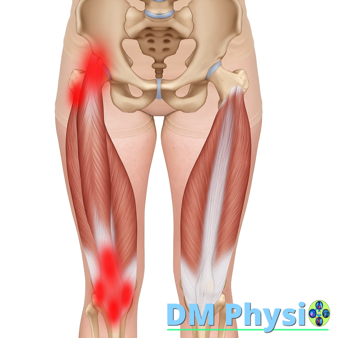
Anatomy and grip
It is located on the front of the thigh. Its tendon connects to the knee cap and through it to the tibia. One of the heads (rectus femoris) also starts from the pelvis.
It is often overloaded at long sitting with straight knees and lots of squats without active glutes.
The location of the pain is shown in red
- Pain in front of thigh and around the kneecap (above/below).
- Intensifies when climbing/descending stairs, squatting and long standing with "locked" knees.
- Tenderness to pressure on cap tendon, morning stiffness after long sitting.
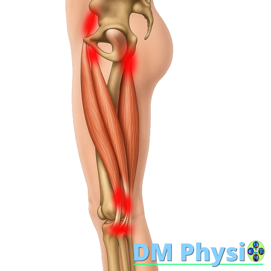
Hamstrings
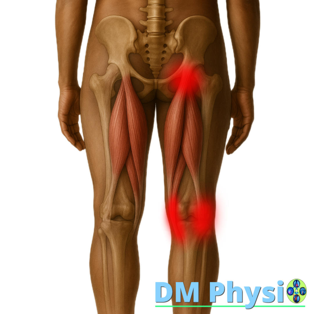
Anatomy and grip
They are located at the back of the thigh – start from the ischium and attach around the knee.
Sitting often inhibits them and makes them inactive; their weakness comes mainly from sitting on them for a long time.
The location of the pain is shown in red
- Pain under the buttock or behind the knee.
- During inactivity, the pelvis rotates forward, which leads to hyperlordosis and additional tension in the lower back.
- It is provoked by bending over, walking faster, standing up from a squat, and sudden accelerations.
Adductors (inner thigh)
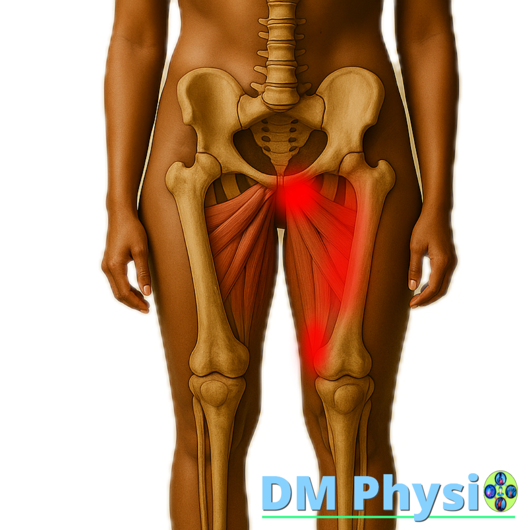
Anatomy and grip
Group of muscles by inner thigh, which connect the pelvis to the femur.
They are often overloaded at wide stride, sharp lateral movements (football, dancing) and long sitting with crossed legs.
The location of the pain is shown in red
- Pain in inner thigh, groin or medially around the knee.
- It gets worse when squatting, standing up, taking a wide stride, or walking for a long time.
- A "pulling" sensation when bringing the knees together or pressing a pillow between them.
TFL / Iliotibial Band (outer thigh)
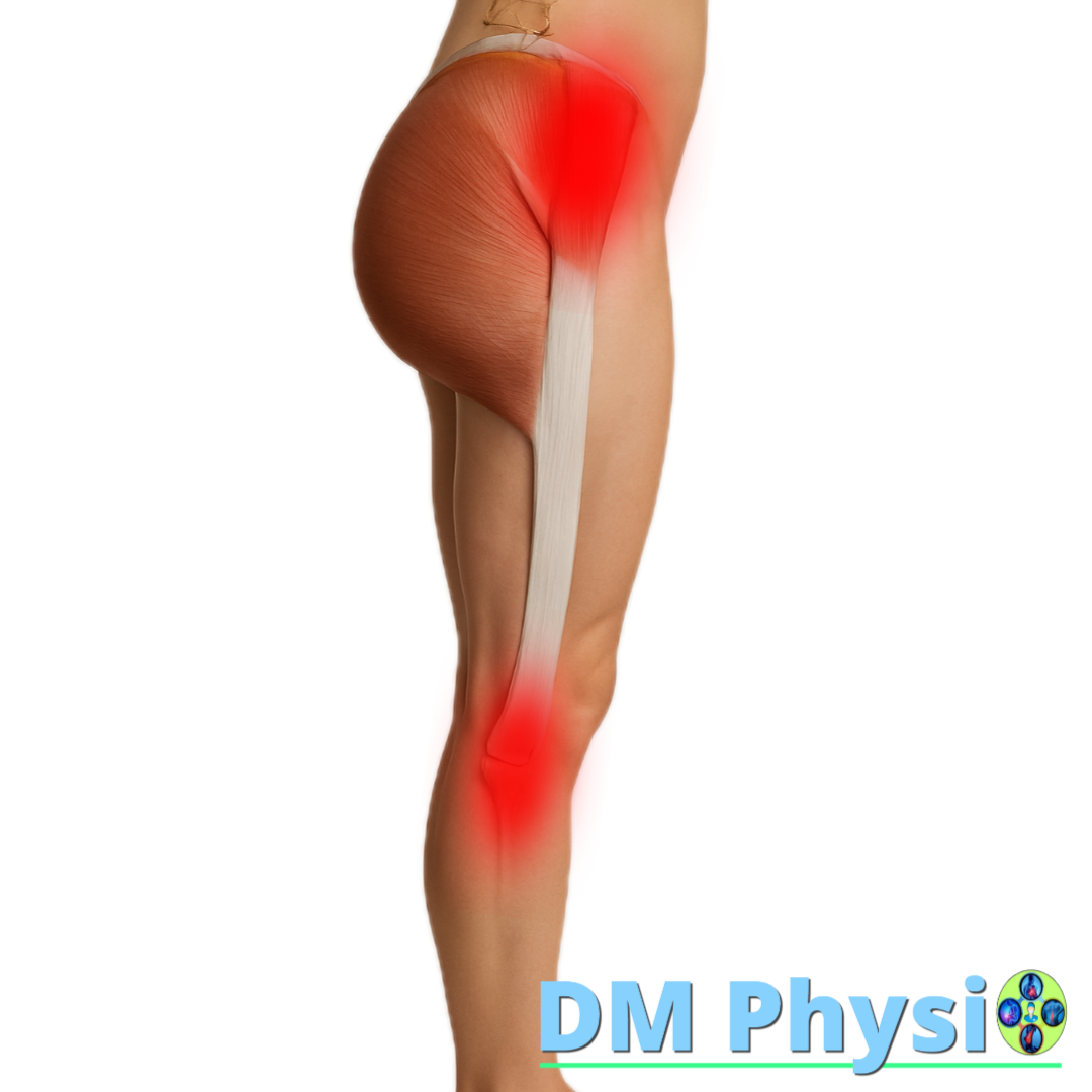
Anatomy and grip
A small muscle front-outside thigh, which passes into a broad fascia (IT band) reaching the knee.
It often "takes over" work with weak buttock stabilizers and strains when sitting for a long time with a leg at the sides.
The location of the pain is shown in red
- Pain on the outer side of the thigh and on the outside of the knee.
- It is triggered by stairs, running down/incline and standing on one leg for a long time.
- Sometimes a "clicking" sensation in the outer knee when squatting/standing.
m. popliteus (behind the knee)

Anatomy and grip
A small, deep muscle behind the knee, which "unlocks" the knee from a fully extended position.
Sensitive to the habit of standing with hyperextension ("locked" knees) and to small "twists" when stepping crookedly.
The location of the pain is shown in red
- Deep pain behind the knee, a "prick" when going down stairs or turning sharply.
- Often after long standing with extended knees or a small step aside crookedly.
- A feeling of slight instability when going down an incline.
Calf (Triceps surae)
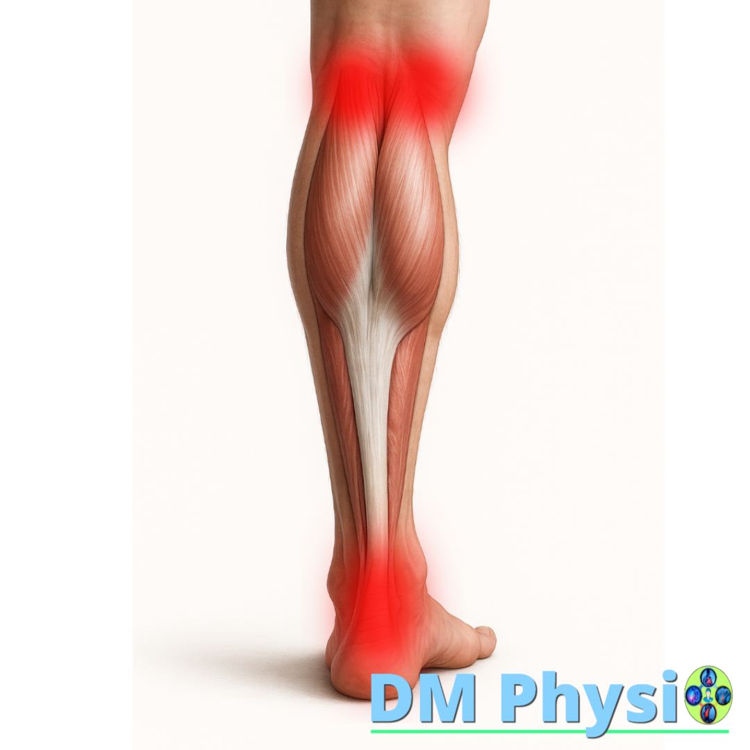
Anatomy and function
The calf consists of gastrocnemius and soleus. Both muscles are captured through Achilles tendon for the fifth and the gastrocnemius also passes through the knee joint. When they are shortened, they limit the movement of the ankle (dorsiflexion) and transfer load to the knee.
Pain and symptoms
- Pain or tightness in the calf, often spreading to heel and achilles.
- Pain behind the knee, which can be either from the inner one, as well as from the outer one country.
- Symptoms increase when climbing stairs, walking on tiptoes and prolonged standing.
- They are often observed morning stiffness of Achilles and night cramps.
m. tibialis anterior, m. extensor digitorum longus and m. extensor hallucis longus (anterior lower leg)
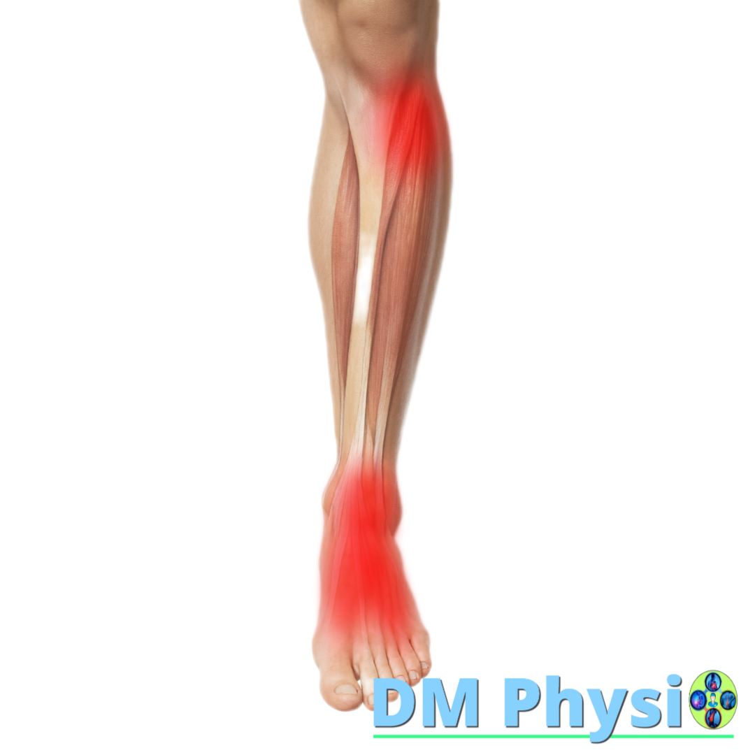
Anatomy and grip
The three muscles are located on the front of the lower leg. m. tibialis anterior lifts the foot and supports the arch, m. extensor digitorum longus extends fingers II–V and assists in dorsiflexion, a m. extensor hallucis longus extends the big toe and also participates in raising the foot.
The location of the pain is shown in red
- Pain and burning externally under the knee and in the front of the ankle.
- Tension and discomfort the back of the foot and the base of the big toe.
- Inflammation and irritation are often provoked by tight shoes or socks.
Fibularis (peroneus) - outer lower leg
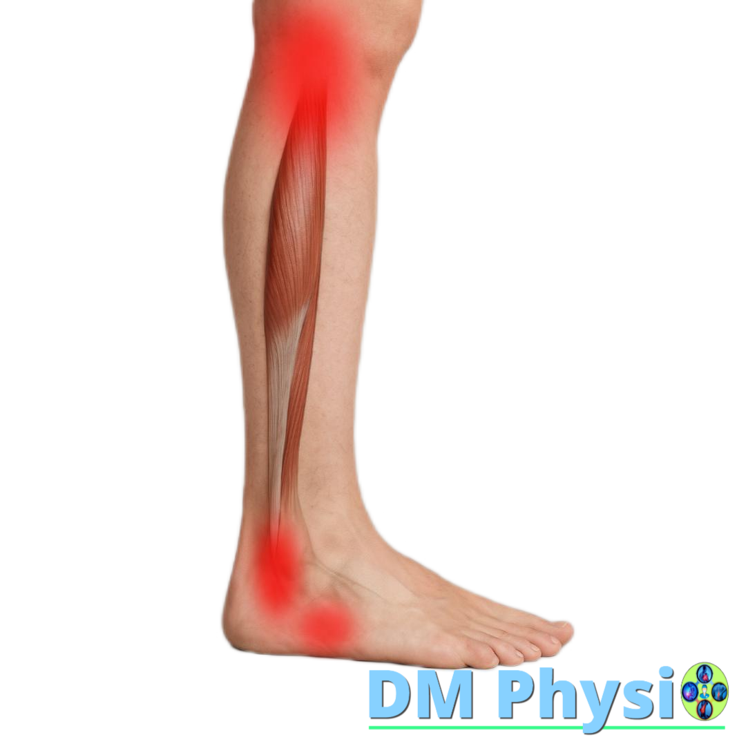
Anatomy and function
Muscles on the outside of the lower leg, which perform eversion of the foot and maintain stability of the ankle and arch of the foot. They protect against sprains and guide the movement of the knee correctly when walking.
The location of the pain is shown in red
- Pain in the outer lower leg, the side of the knee, ankle and foot.
- It is aggravated when walking on uneven ground or when twisting the ankle.
Pes anserinus ("duck's foot" - inside below the knee)
Anatomy and grip
Pes anserinus is general tendon entrapment on three muscles – sartorius, gracilis and semitendinosus. They start from different parts of the pelvis and thigh, but come together and attach on the inside of the tibia (below the knee). There is often a small bursa between the tendon and the bone that reduces friction.
The location of the pain is shown in red
- The pain is often local and burning, felt below and on the inside of the knee.
- In case of trauma or overload of any of the three muscles, the pain can be felt both in the area of the pes anserinus, as well in the other place of capture of the corresponding muscle (eg in the pelvis or thigh).
- It is provoked by going down stairs, squatting and prolonged walking.
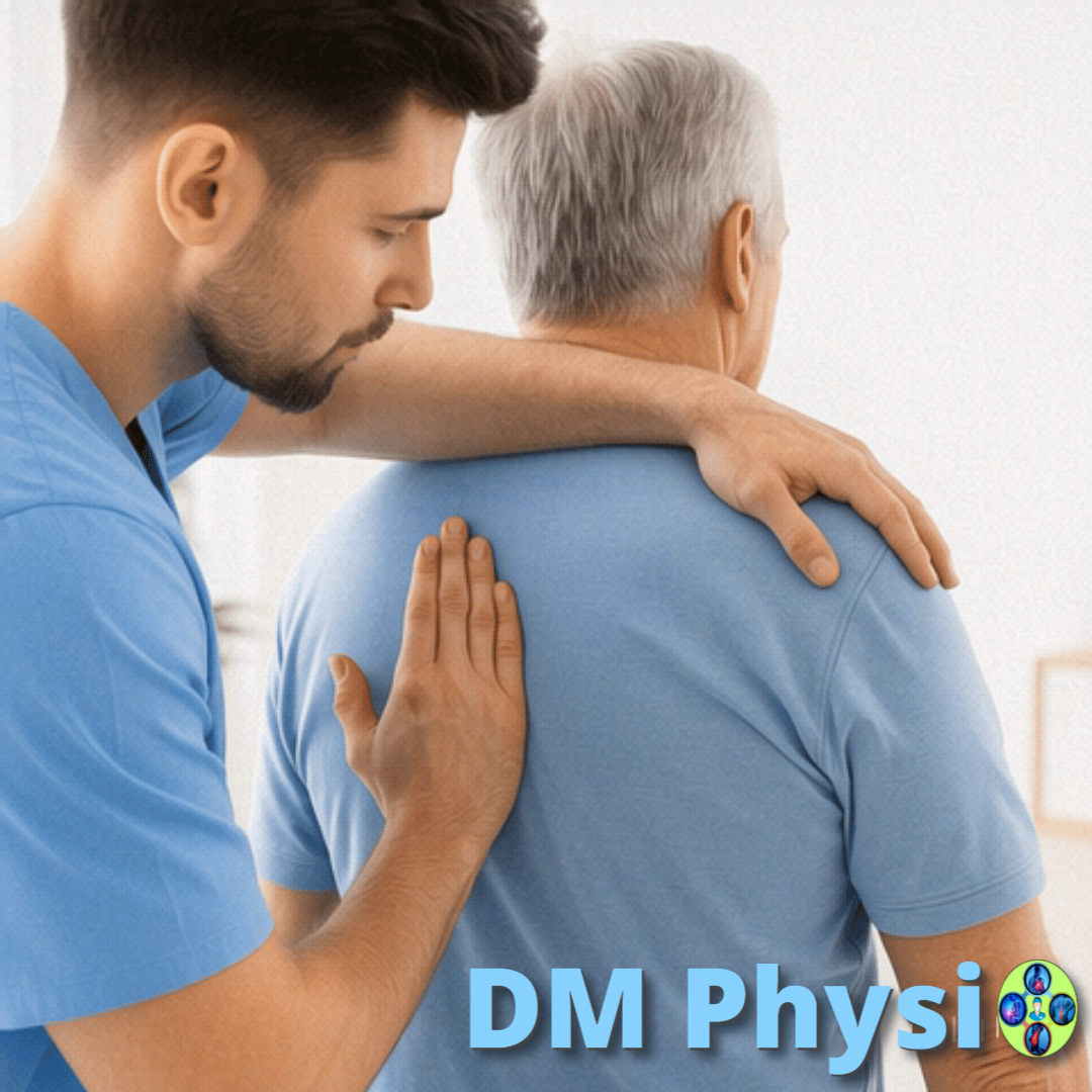
How to deal with hip and knee pain?
We focus on relaxation, rebalancing and changing habits.
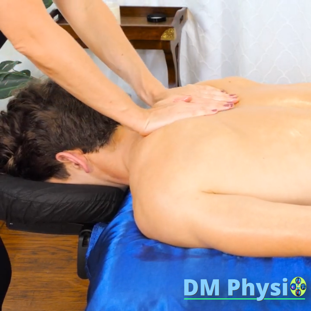
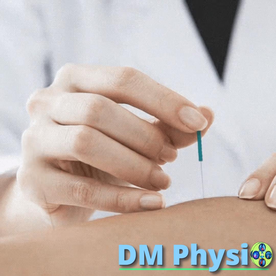
1. Relaxation of tense muscles
Deep tissue massage, myofascial release and manual therapy for anterior/inner/outer/posterior thigh, area behind the knee, calf and lower leg - for rapid pain reduction and release of movement.
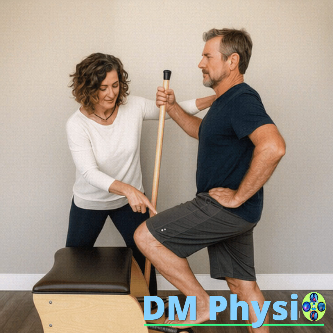
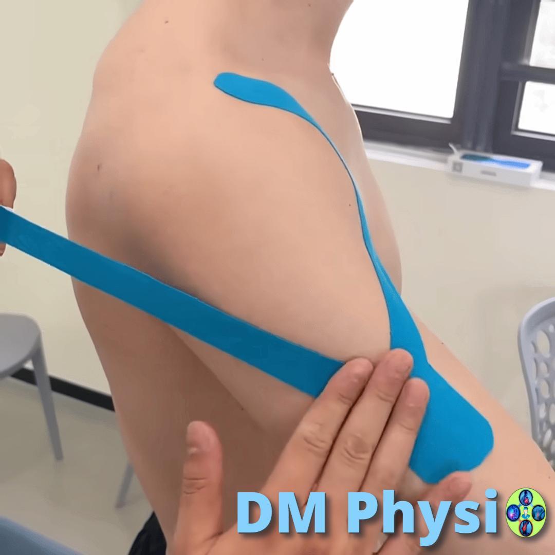
2. Strengthening and balancing
Stretching shortened and strengthening weak muscles. We teach "soft knee" in standing, smooth folding when stepping and a stable arch of the foot to distribute the load evenly.
3. Correction and prevention
Practical tips for sitting, standing, walking and sleeping: take frequent breaks from sitting, avoiding "locked" knees, appropriate footwear and dosage of the load. So we prevent the pain from returning.