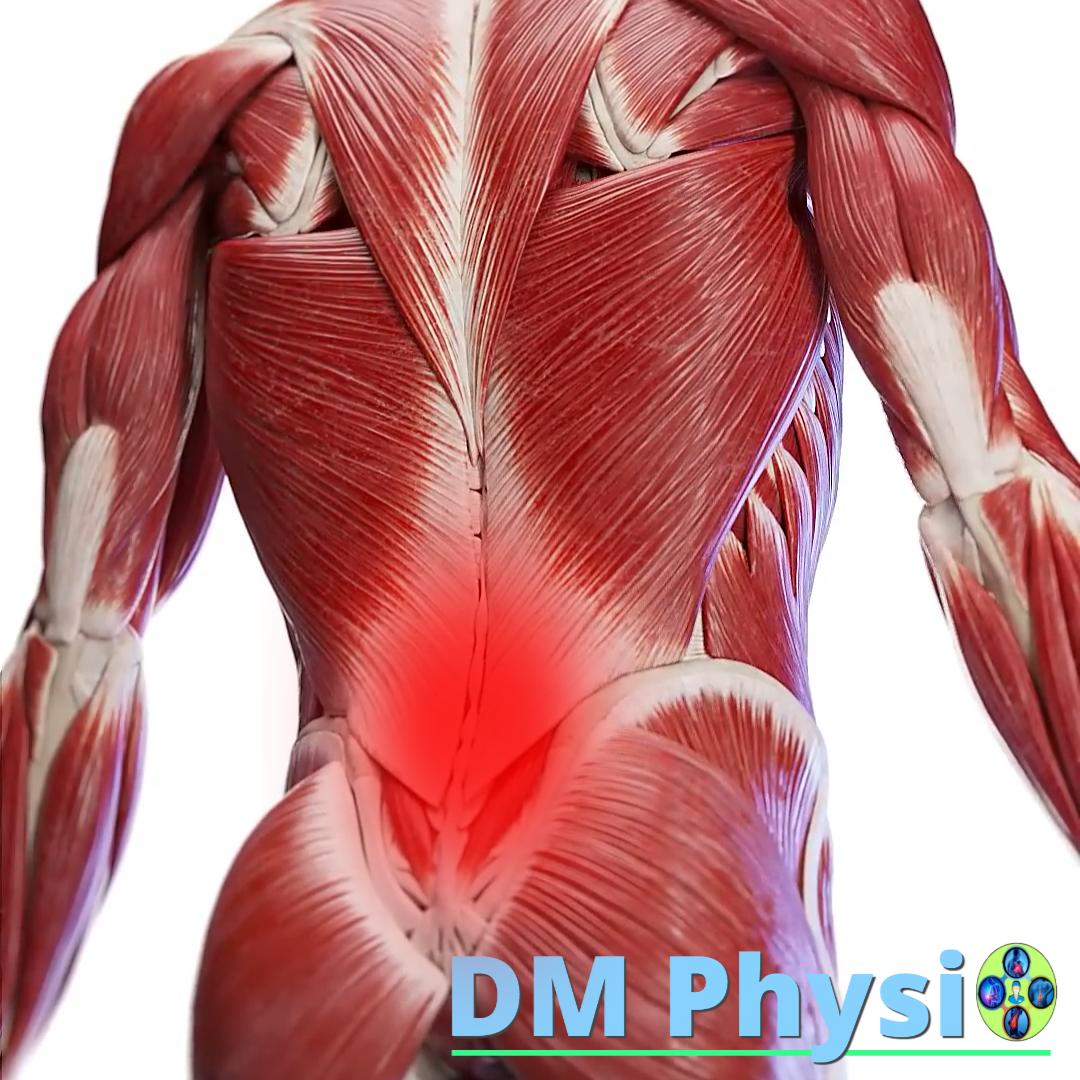
Muscular pain in the lower back
Find out the main causes of lower back pain and what can help.
Why does the lower back hurt?
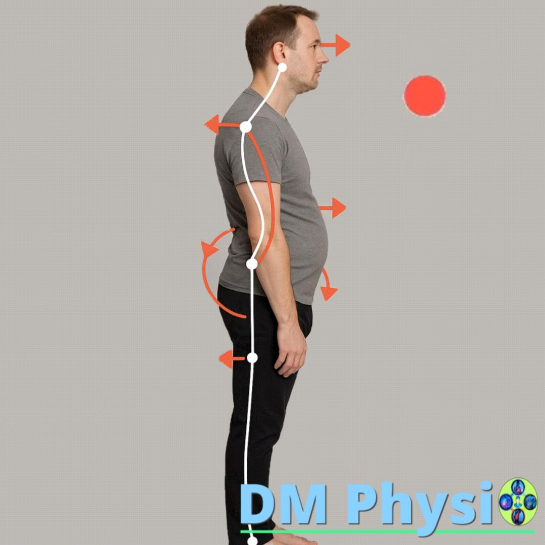
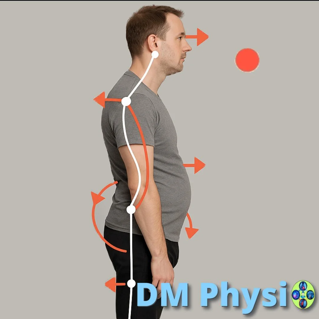
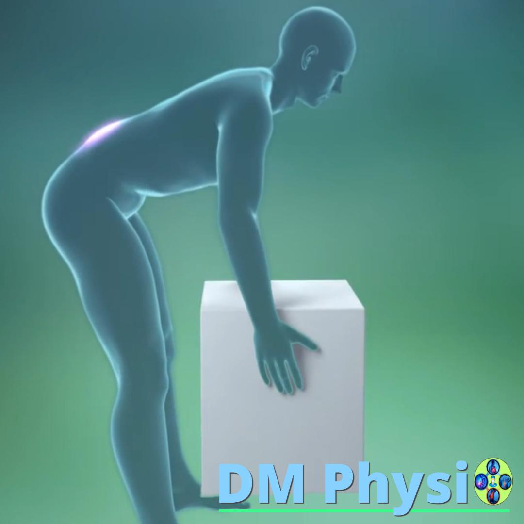
Low back pain is often caused by prolonged sitting, standing or sleeping in poor positions, which puts strain on the lumbar region. This happens because the muscles that are responsible for maintaining proper posture do not function effectively because they have been overstretched or squeezed for a long time without being activated.
When there are weak or inactive muscles, the body compensates for this weakness by stressing other muscles. This is the beginning of a muscle imbalance that leads to a change in posture and body position, and ultimately to chronic low back pain.
How is muscle imbalance formed?
Improper posture
Prolonged sitting, poor posture or lack of movement lead to stretched or tight muscles that lose their strength and ability to support the body.
Compensation
To compensate for the weakness of inactive muscles, the body transfers the load to other muscles. They overwork, become hypertensive (excessively tense) and develop microtraumas.
Muscle spasm and pain
Overworked muscles react with spasm and the formation of trigger points. These points cause sharp or dull pain, restrict movement, and can radiate pain to other parts of the body, creating a vicious cycle.
Which muscles are most commonly affected?
m. quadratus lumborum (Square muscle of the lower back)
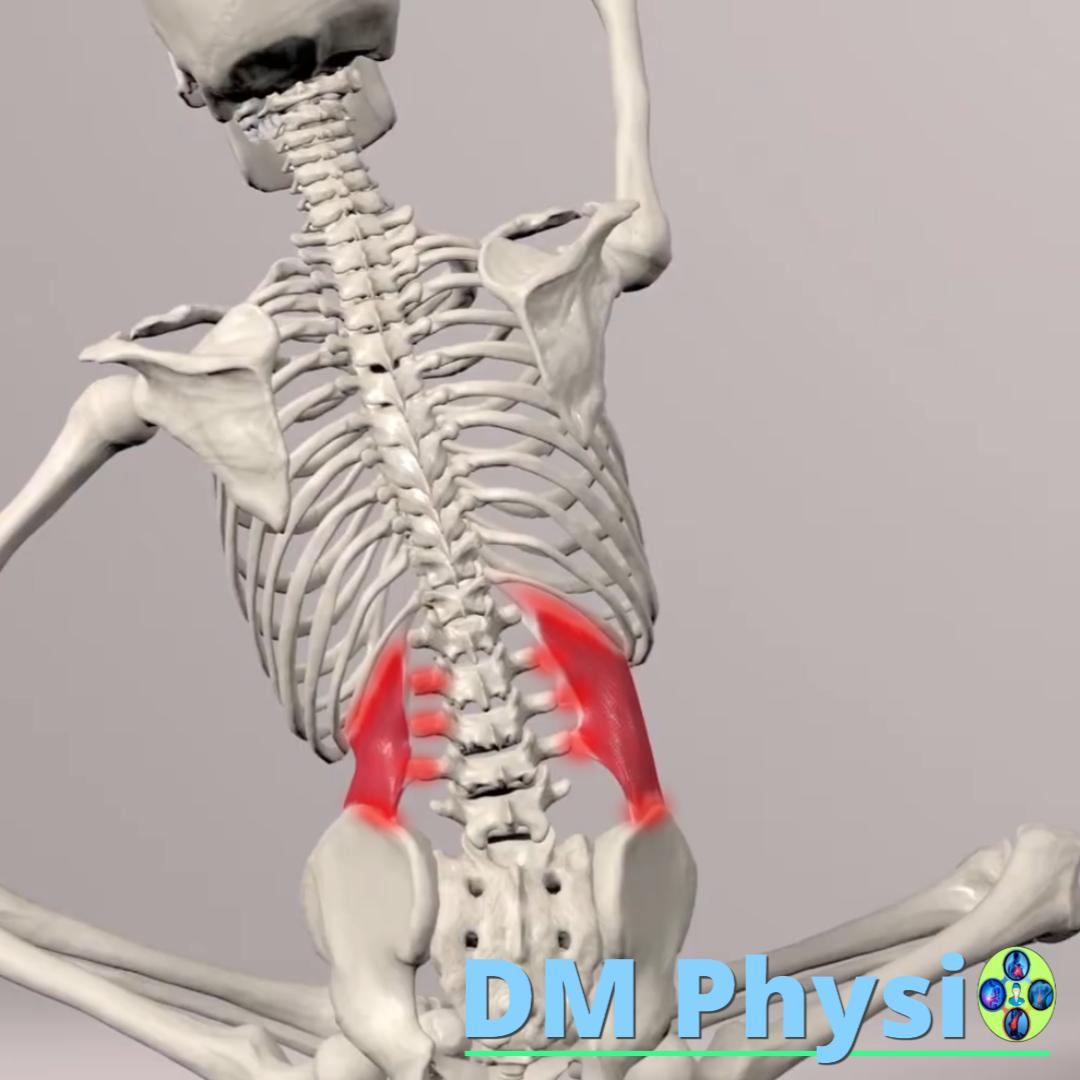
Anatomy and grip
A deep muscle located in lower back. Connects the 12th rib to the lumbar vertebrae and pelvis. It is one of the main sources of low back pain as it is a key stabilizer of the lumbar spine.
The location of the pain is shown in red
- Deep, severe pain in the lower back, which is also felt when coughing or sneezing.
- The pain may radiate to the buttock, thigh, or groin, often mimicking other conditions.
- Stiffness and stiffness that interfere with standing.
m. iliopsoas (Iliopsoas muscle)
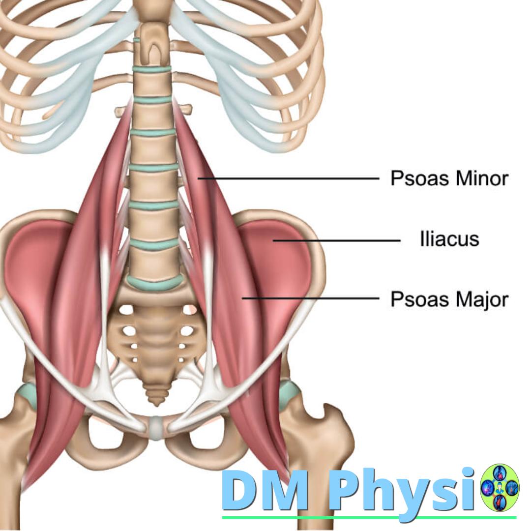
Anatomy and grip
The iliopsoas muscle is a combination of two muscles: psoas major and iliacus. They fuse and attach to the lesser trochanter of the femur. This muscle is key to hip flexion.
The location of the pain is shown in red
- Deep, dull pain in front of thigh, which may spread to the lower back or groin.
- Pain on standing, especially when trying to stand up after sitting for a long time.
- Pain when walking, especially when lifting your leg up.
m. erector spinae (Erecting muscles of the spine)
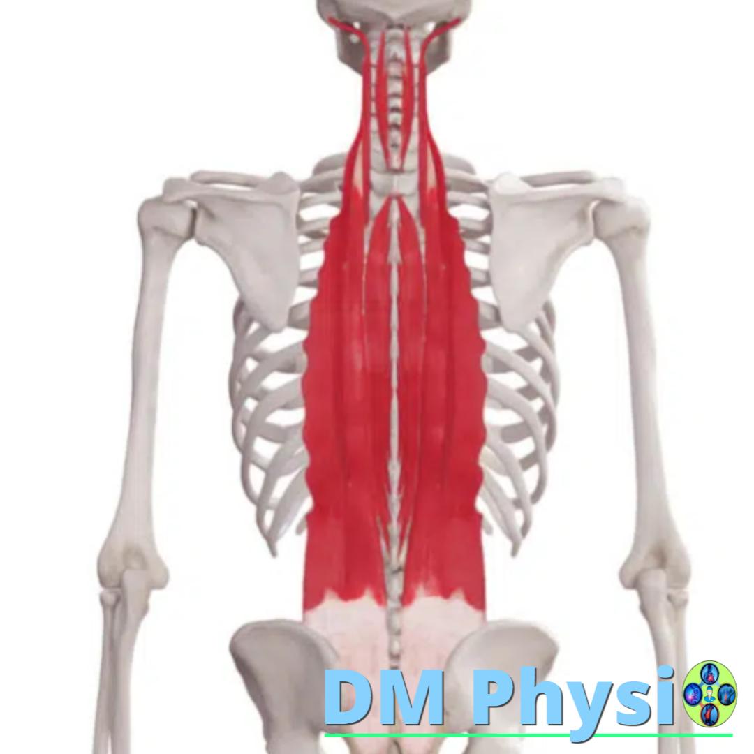
Anatomy and grip
They consist of three muscular columns that extend along the entire spine. They are responsible for maintaining upright posture and are particularly vulnerable in the lower part (lumbar region) where they are often overworked.
The location of the pain is shown in red
- A dull, constant pain, located on the side of the spine.
- It may feel like a "tightness" or "stiffness" in the lower back.
- The pain may spread to the buttock or abdomen.
m. gluteus medius/minimus
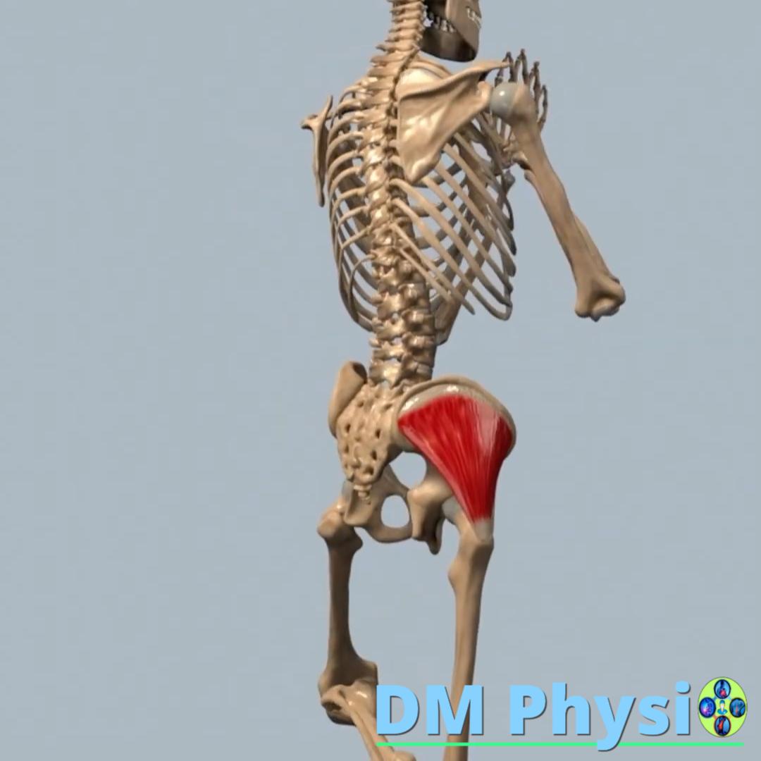
Anatomy and grip
They are located deep under the gluteus maximus muscle and connect the pelvis with the femur. They are critical for pelvic stability. Weakness in these muscles leads to instability and excessive stress on the lower back muscles.
The location of the pain is shown in red
- Deep pain in buttock, which can spread to the waist, thigh and even below the knee, mimicking sciatica.
- The pain worsens with prolonged sitting or standing.
- A feeling of weakness or unsteadiness in the leg.
m. gluteus maximus
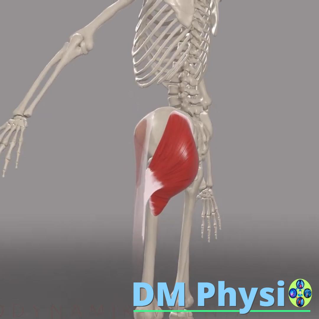
Anatomy and grip
The largest muscle in the body that forms the main part of the buttock. It connects the pelvis with the femur and has a key role in stabilizing the pelvis and lower spine.
The location of the pain is shown in red
- A dull, deep pain in the buttock, which can be felt when sitting.
- The pain may radiate to the sacroiliac joint or lower back.
- It can cause pain along the entire length of the back of the thigh.
m. piriformis
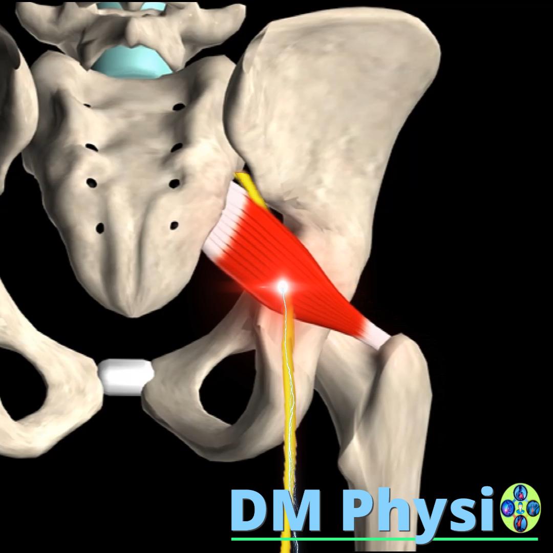
Anatomy and grip
A small, deep muscle located in the buttock. Connects sacrum with the top of the femur. The sciatic nerve runs very close to it. When this muscle shortens or tightens, it can press on the nerve, causing severe pain.
The location of the pain is shown in red
- Sharp pain in the buttock, which may resemble sciatica (piriformis syndrome).
- Radiating pain down back of thigh and calf, sometimes down to foot.
- Pain when sitting or driving for long periods of time.
m. latissimus dorsi (Broad back muscle)
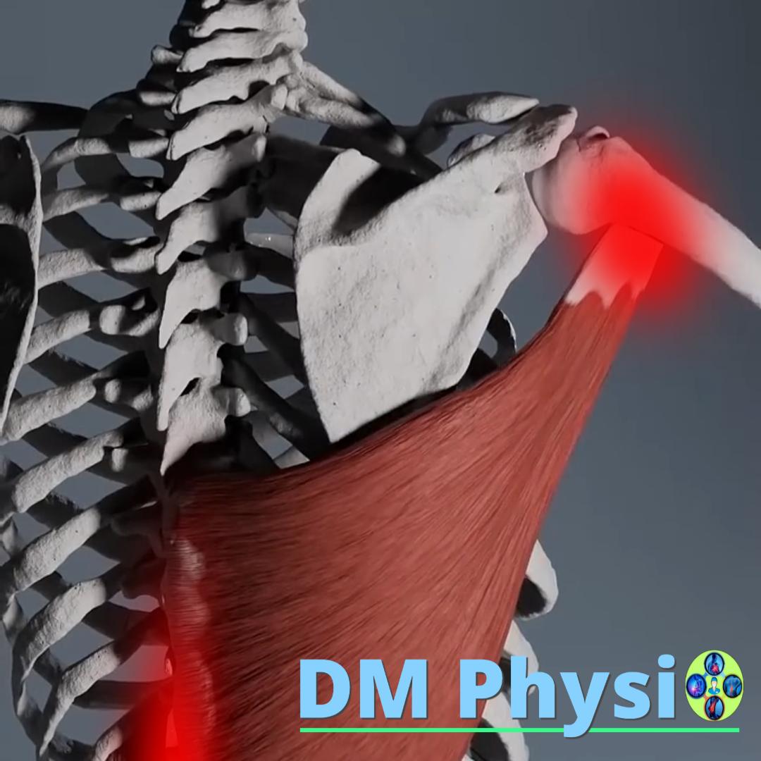
Anatomy and grip
The largest muscle in the upper body, located in the lower back. It attaches to the spine, pelvic bone and attaches to the upper arm bone. Due to its wide range, it has a strong influence on the stability and movement of the spine.
The location of the pain is shown in red
- Lower back pain, which can spread to the shoulder, inner arm, and even the elbow.
- The pain increases with movements of the arms above the head or with rotation of the torso, which is typical of problems with this muscle.
- It is often confused with kidney pain or shoulder pain, but its origin is muscular.
m. transversus abdominis (Transverse abdominal muscle)
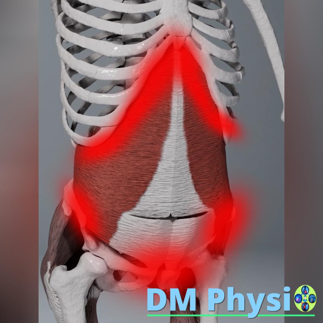
Anatomy and grip
The deepest abdominal muscle that wraps around the torso like natural "corset" of the body. Its main role is to stabilize the spine in front. Weakness in this muscle can lead to a lack of support and strain on the muscles in the lower back.
The location of the pain is shown in red
- A deep, dull ache in the lower abdomen or groin area.
- It often feels like "tightness" or "pulling" in the abdominal area.
- Weakness in the muscle can contribute to low back pain because its role is key to core stability.
m. obliquus externus abdominis (External oblique abdominal muscle)
Anatomy and grip
The largest and most superficial of the three lateral abdominal muscles, located in the side of the abdomen. It is important for the rotation of the body, but an imbalance in it can lead to overstrain in the lower back muscles.
The location of the pain is shown in red
- Pain in the groin area and around the front of the pelvis, often confused with a hernia.
- Pain in the lower abdomen and on the side of the navel.
- A deep, dull ache that worsens with movement of the torso.
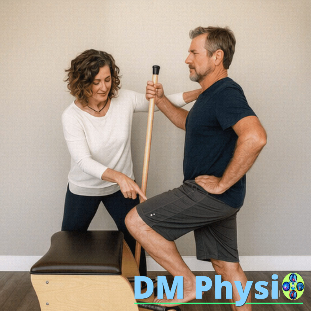
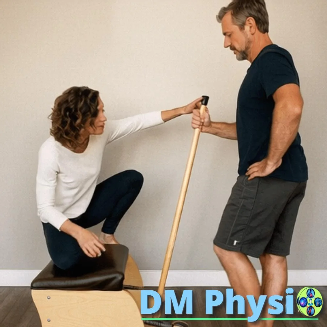
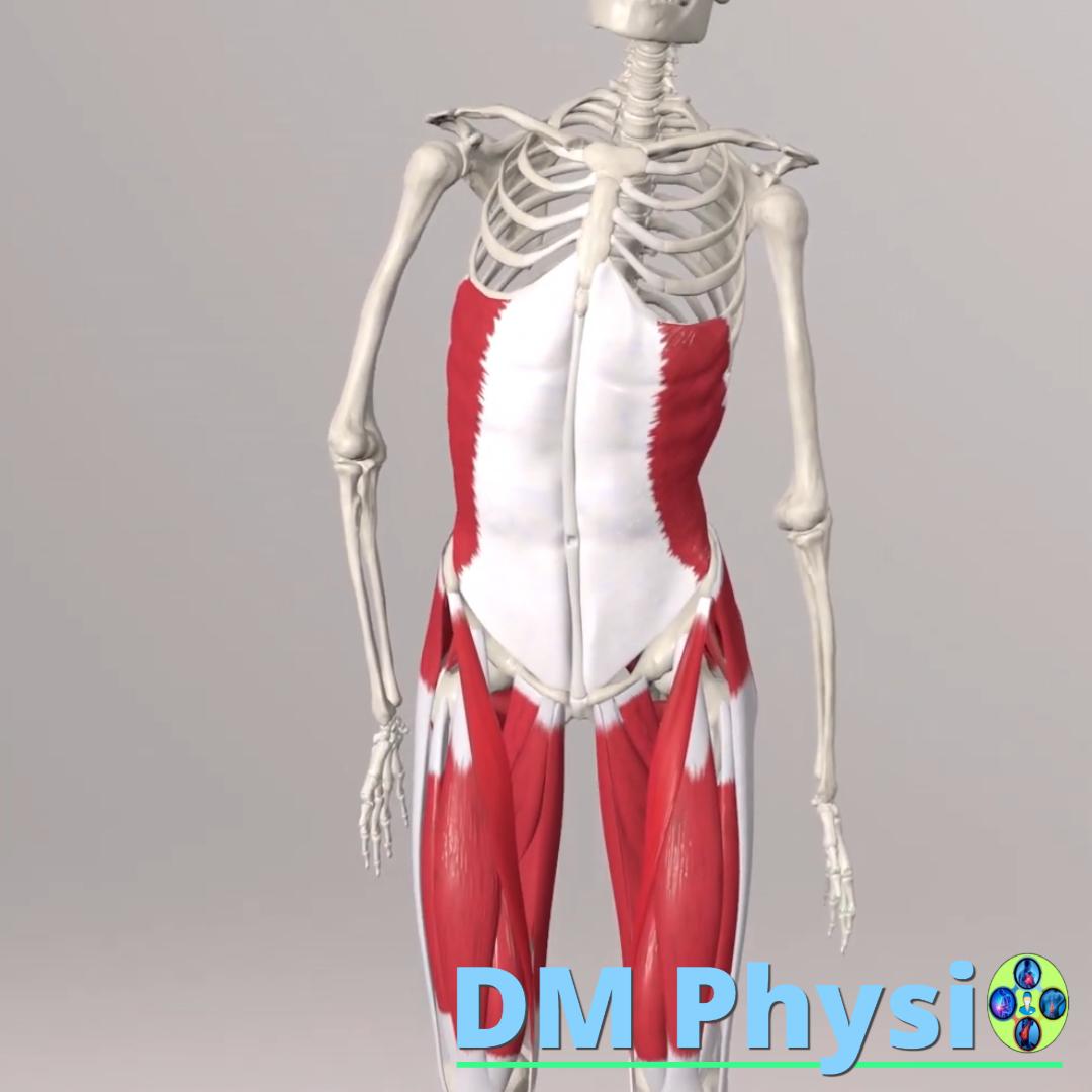
How to deal with low back pain?
To reduce pain, we focus on relaxing, restoring and strengthening the muscles of the lower back.
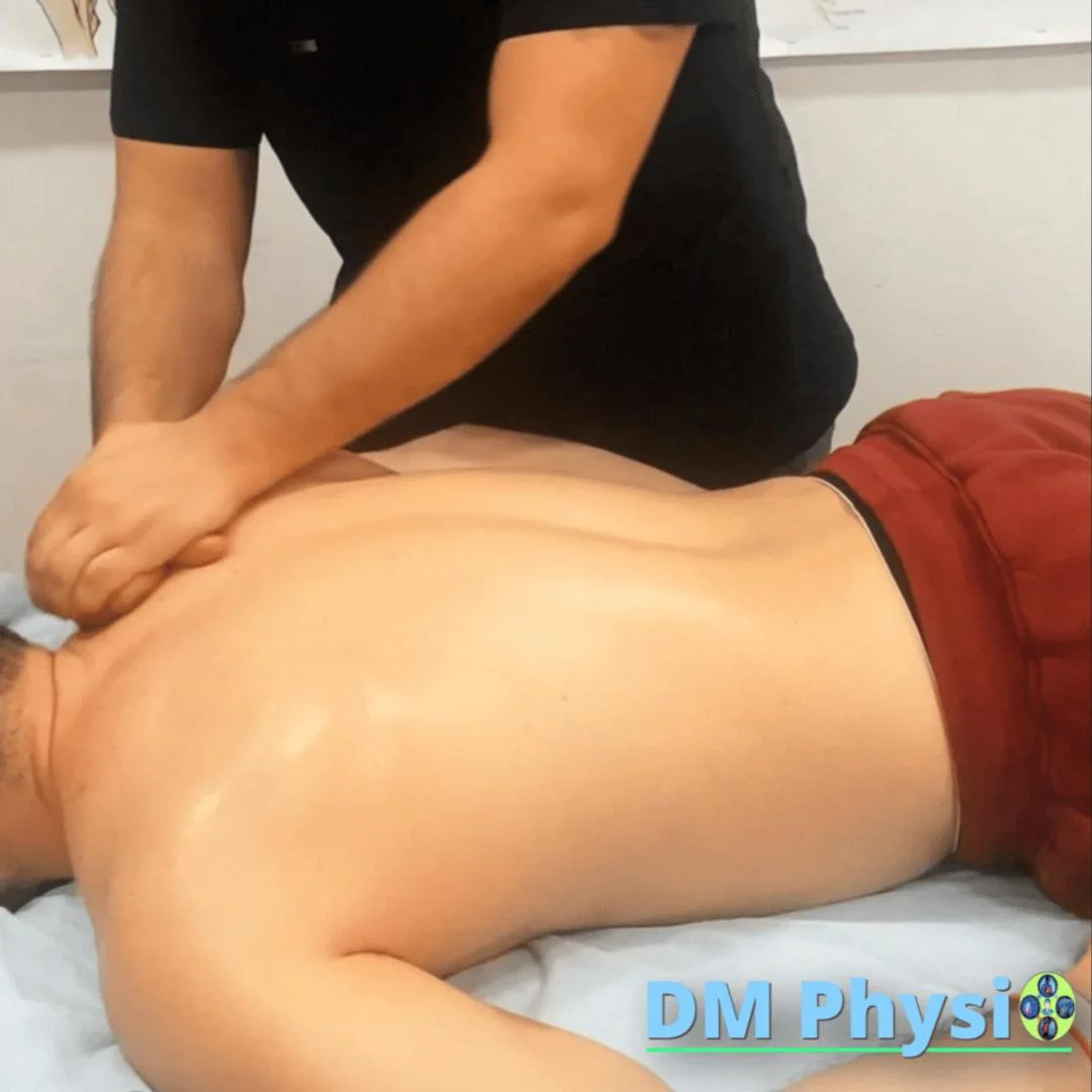
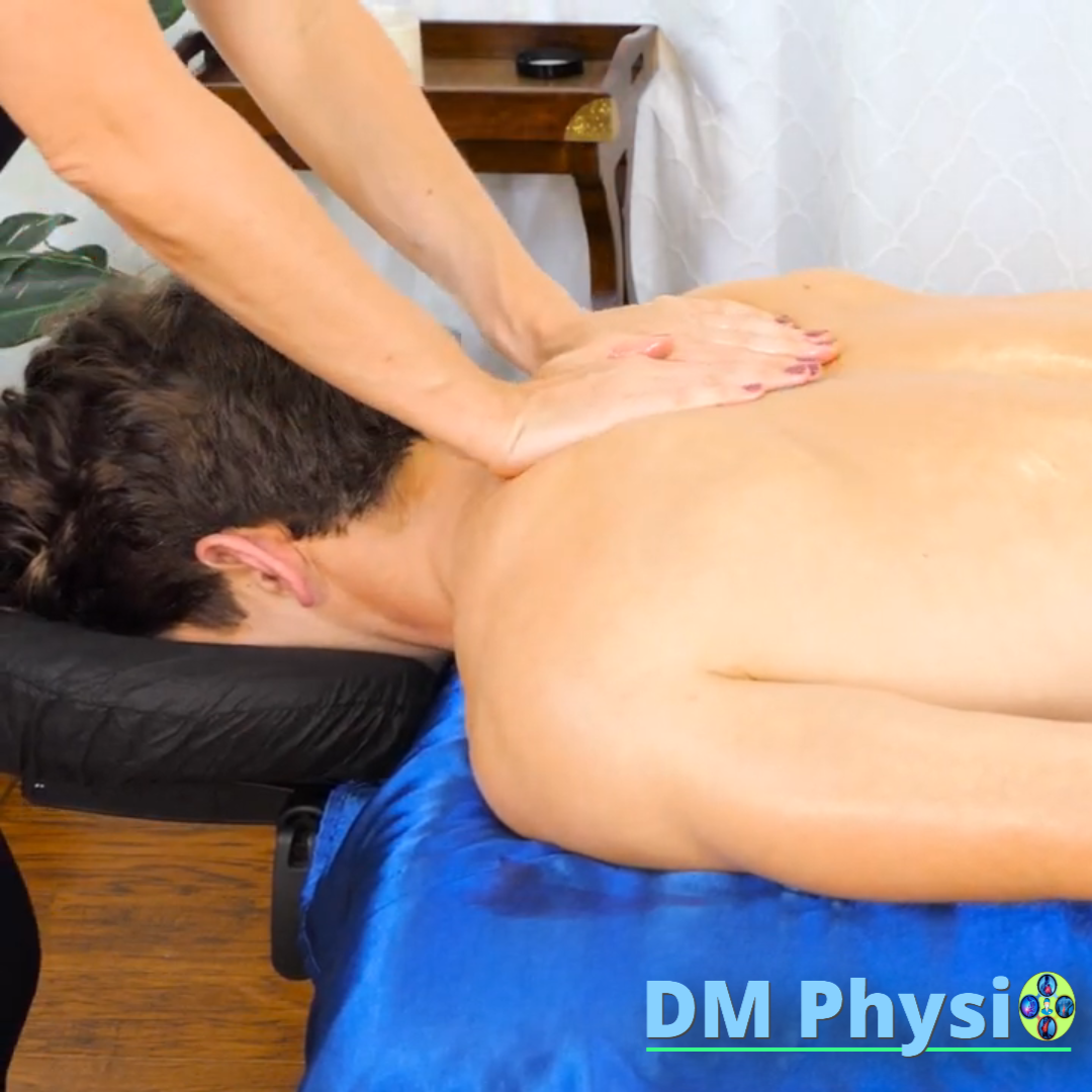
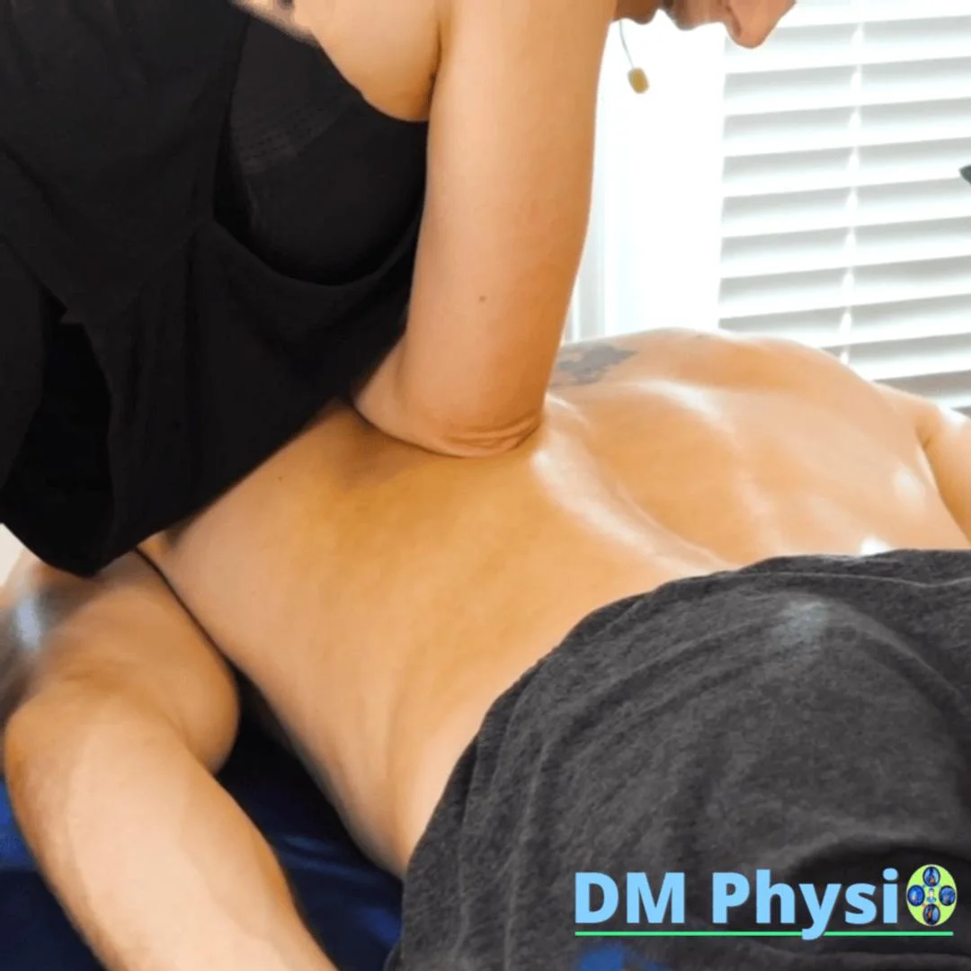
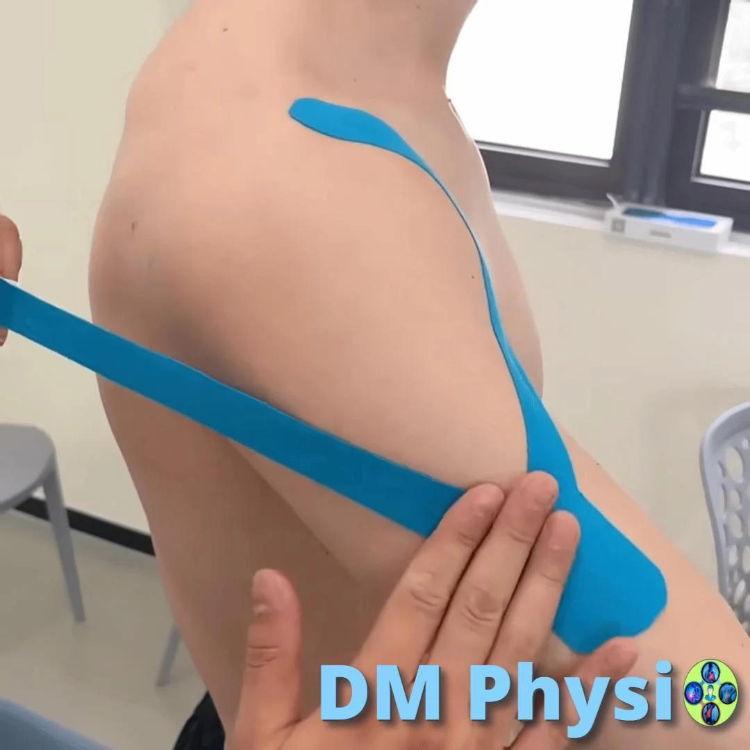
1. Relaxation of tense muscles
Through techniques such as deep tissue massage, myofascial release and manual therapy, we relax contracted muscle fibers and trigger points, to support better function and reduce pain.
2. Strengthening and balancing
Once the muscles are relaxed, the focus is on strengthening the abdominal wall and gluteal muscles, which are key to low back and pelvic stability.
3. Correction and prevention
We teach you specific exercises and tips for correct ergonomics and posture, to prevent future pain and keep your lower back healthy and mobile in the long term.