Muscular pains in the chest and abdomen
Understand how muscle tension, loading and movement control can contribute to chest or abdominal discomfort.

Why does this pain occur?
Chest and abdominal pain can be caused by overstrain of the respiratory muscles, intercostal muscles, or spasms of the abdominal muscles. It is often confused with heart problems or problems with internal organs, but in many cases it is related to muscle dysfunctions. In a number of situations, the cause is precisely trauma - for example, after a fall or a direct blow to the area, which leads to tension or spasm of the affected muscles.
Muscle pain in the chest often feels like a stabbing pain that gets worse with breathing or movement. Correct diagnosis by a specialist is key.
How does muscle pain develop?
Chest and abdominal pain rarely comes on "suddenly." It usually accumulates as a result of habits, muscle imbalances and limited rib/thoracic and pelvic mobility. Here is the most common sequence:
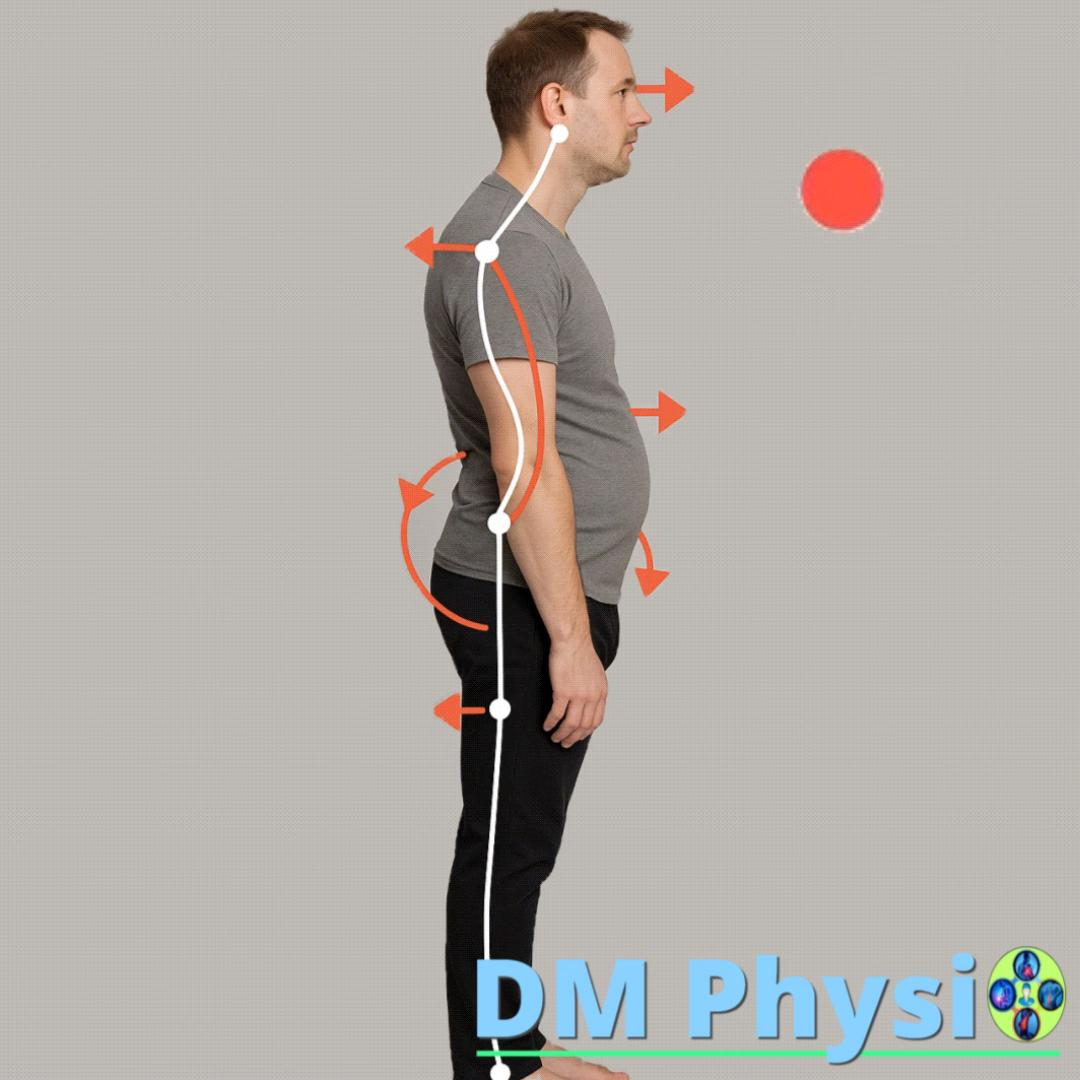
Improper postures and overexertion
Prolonged sitting, bent shoulders, training without technique or unilateral loads lead to increased tension in the intercostal, chest and abdominal muscles.
Muscle imbalance
Some of the muscles shorten and become overactive, while their antagonists weaken. This displaces the ribs and shoulder girdle.
Limited mobility and shallow breathing
Reduced mobility of the ribs and ribcage-related changes the breathing pattern. During shallow breathing, the intercostal and auxiliary respiratory muscles are overstretched.
Which muscles cause pain?
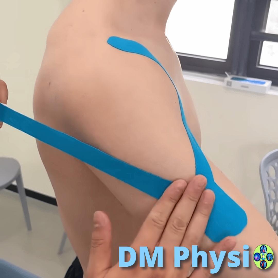
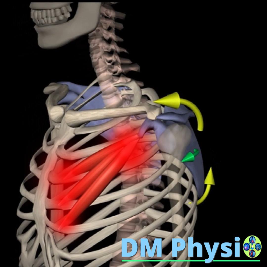
m. serratus anterior (serratus anterior)
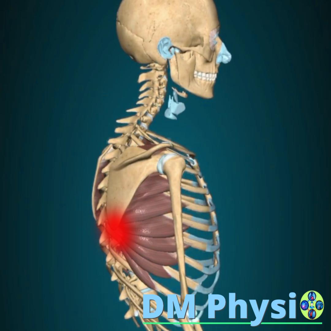
Anatomy and grip
It is located on the side of the chest, under the armpit. It starts from the first 8-9 ribs and attaches to the inner edge of the scapula.
The location of the pain is shown in red
- A stabbing pain in the chest, especially when breathing deeply or coughing.
- Pain in the side of the chest.
- It can feel like a "strange" pain under the breast.
m. rectus abdominis (rectus abdominis)
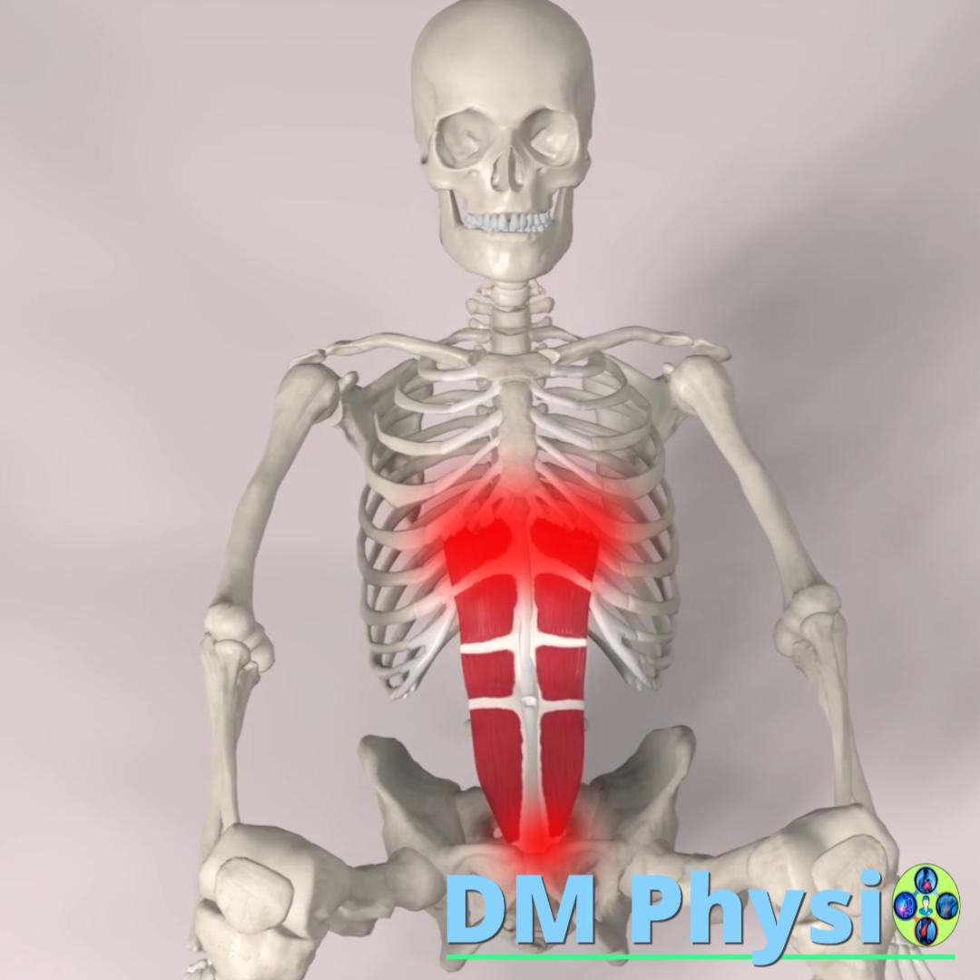
Anatomy and grip
This muscle is located along the front of the abdomen. It starts from the pelvis and attaches to the lower ribs and sternum.
The location of the pain is shown in red
- A dull, throbbing pain over the entire abdomen, which increases with tension or movement.
- It can be felt as pain in the internal organs.
- Sensitivity to touch.
m. transversus abdominis (transverse abdominal muscle)
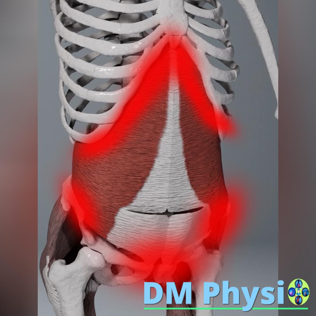
Anatomy and grip
It is located deep below the rest of the abdominal muscles. It starts from the lumbar fascia, pelvis and lower ribs and attaches to the linea alba (white line of the abdomen).
The location of the pain is shown in red
- Deep, pressing pain in the lower abdomen and pelvis.
- It can feel like abdominal cramps or pain related to the pelvic organs.
- It is often associated with lower back problems due to its role as a stabilizer.
m. pectoralis minor (small chest muscle)
Anatomy and grip
It is located under the pectoralis major muscle. It starts from the 3rd, 4th and 5th ribs and attaches to the coracoid process of the scapula.
The location of the pain is shown in red
- Chest pain, shoulder and forearm, often described as burning.
- It can cause numbness and tingling in the fingers, mimicking carpal tunnel syndrome.
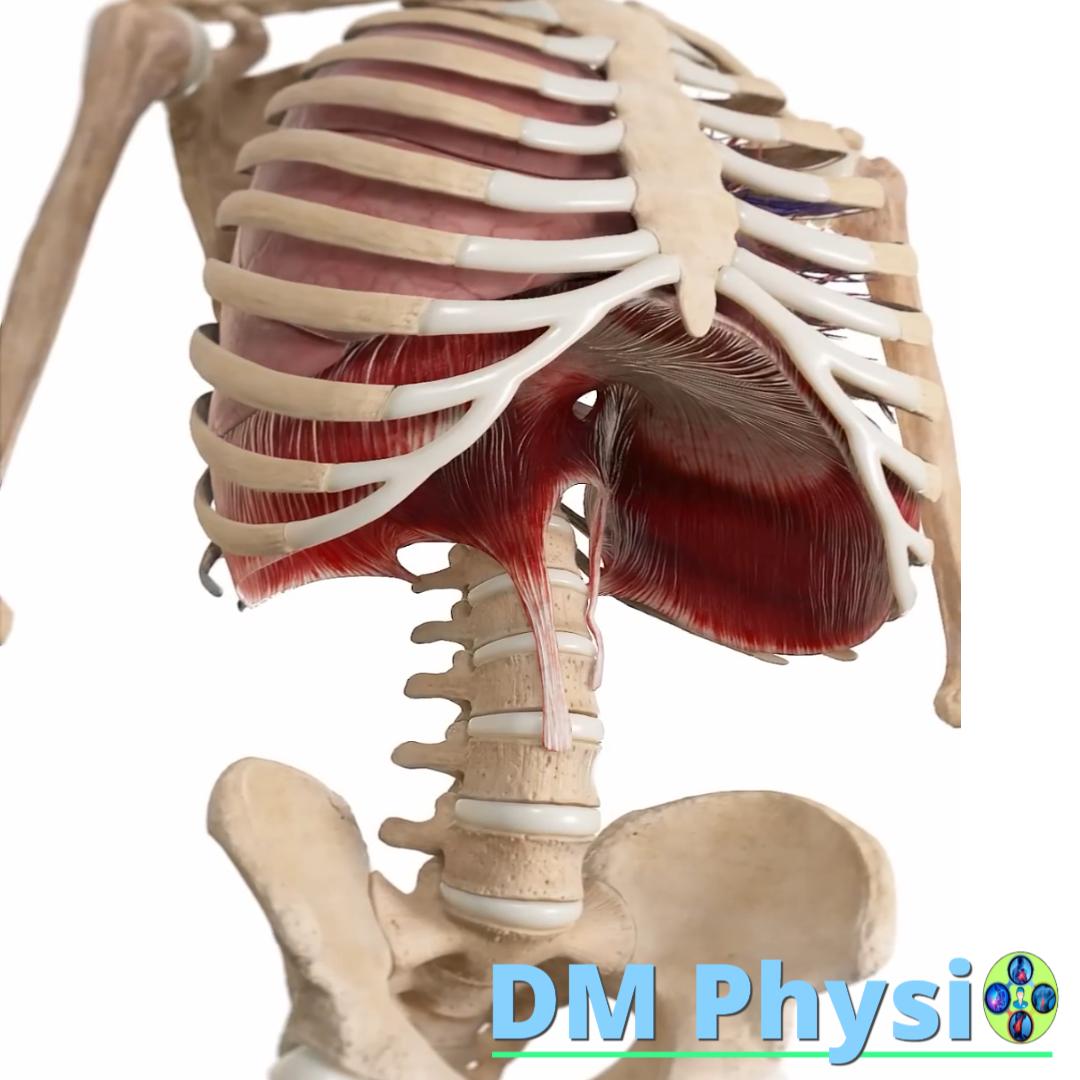
Diaphragm
Anatomy and grip
The diaphragm is the main respiratory muscle that separates the thoracic cavity from the abdominal cavity. It starts from the lower edge of the chest, sternum and lumbar vertebrae, and attaches to the central tendon.
- Chest or abdominal pain, often confused with heart problems or problems with internal organs.
- It can feel like pressure or spasm, especially when taking a deep breath.
- It often occurs after trauma (a fall or a direct blow to the area) or an overload of the respiratory muscles.
m. pectoralis major (large chest muscle)
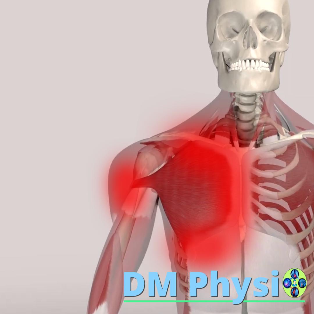
Anatomy and grip
The muscle is located in the front of the chest. It starts at the collarbone, sternum and ribs and attaches to the humerus.
The location of the pain is shown in red
- A deep, dull ache in the chest which may spread to the shoulder, arm and fingers.
- It often mimics symptoms of a heart attack.
- Limitation of movements in the shoulder joint.
How to deal with pain?
Pain is not an enemy – it is a signal that your body needs attention. The solution is a combination of relaxation, recovery and building new healthy habits.
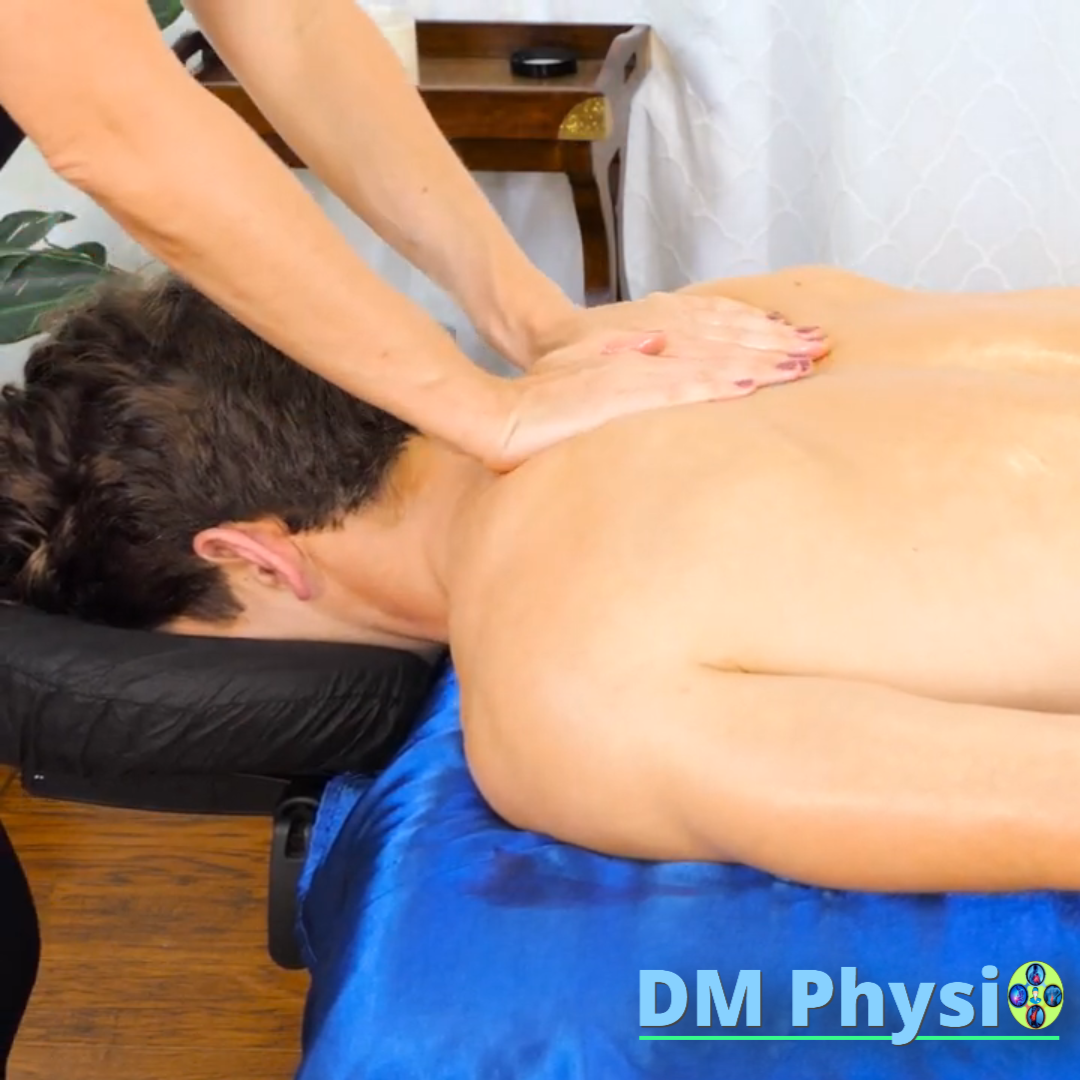
1. Release and release
With the help of manual techniques, massages and mobilization remove accumulated tension, spasms and trigger points. This restores the normal elasticity of the muscles and reduces pain already in the first sessions.
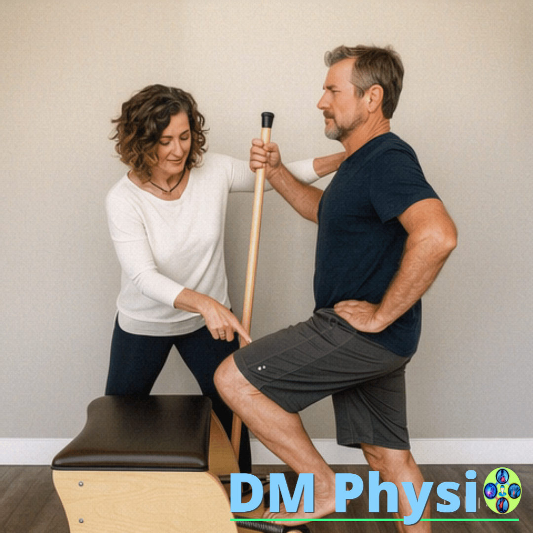
2. Strengthening and balance
The real solution is to put power back where it's missing. We work for activation and strengthening of weak muscles, to restore the balance between different muscle groups and the body can move freely and without tension.
3. Correction and prevention
You learn how to move and maintain correct posture and healthy habits in your daily life. Thus, you not only eliminate the current pain, but also prevent future problems with the spine, joints and muscles.