Muscular pains in lower leg and foot
This page will help you quickly identify which muscle is the likely cause of your lower leg and foot pain by comparing the area of discomfort with the animations shown.
Why do lower legs and feet hurt?
Muscle pain often comes from overload or trauma. Let's take Vesco for example. He hasn't worked out in a few months, but suddenly decides to do it super hard workout. The next day his muscles are literally "on fire". This is inflammation from microtraumas in the muscle fibers - a normal reaction to such a load.
However, when a muscle overloaded, he can not only become inflamed, but yes tighten. In such cases, the muscle does not relax easily and remains tense a long time. This leads to constant discomfort, a feeling of stiffness and pain, which is aggravated by movement or prolonged standing.
When a muscle remains too tight, can form in it the so-called trigger points – small painful nodules. They don't go away easily and can cause problems for years. With each new load, the muscle contracts again, becomes inflamed and the pain returns.
Which muscles most commonly cause lower leg and foot pain?
m. popliteus (behind the knee)

Anatomy and grip
Small, deep muscle of the back of the knee, which "unlocks" the knee from full extension and stabilizes the joint during slight rotations.
Nature of the pain
- Deep pain behind the knee, "poke" when deep squatting or going down stairs
- Provoked after long standing with locked knees or stepping "wrongly"
- A feeling of slight instability when going down an incline
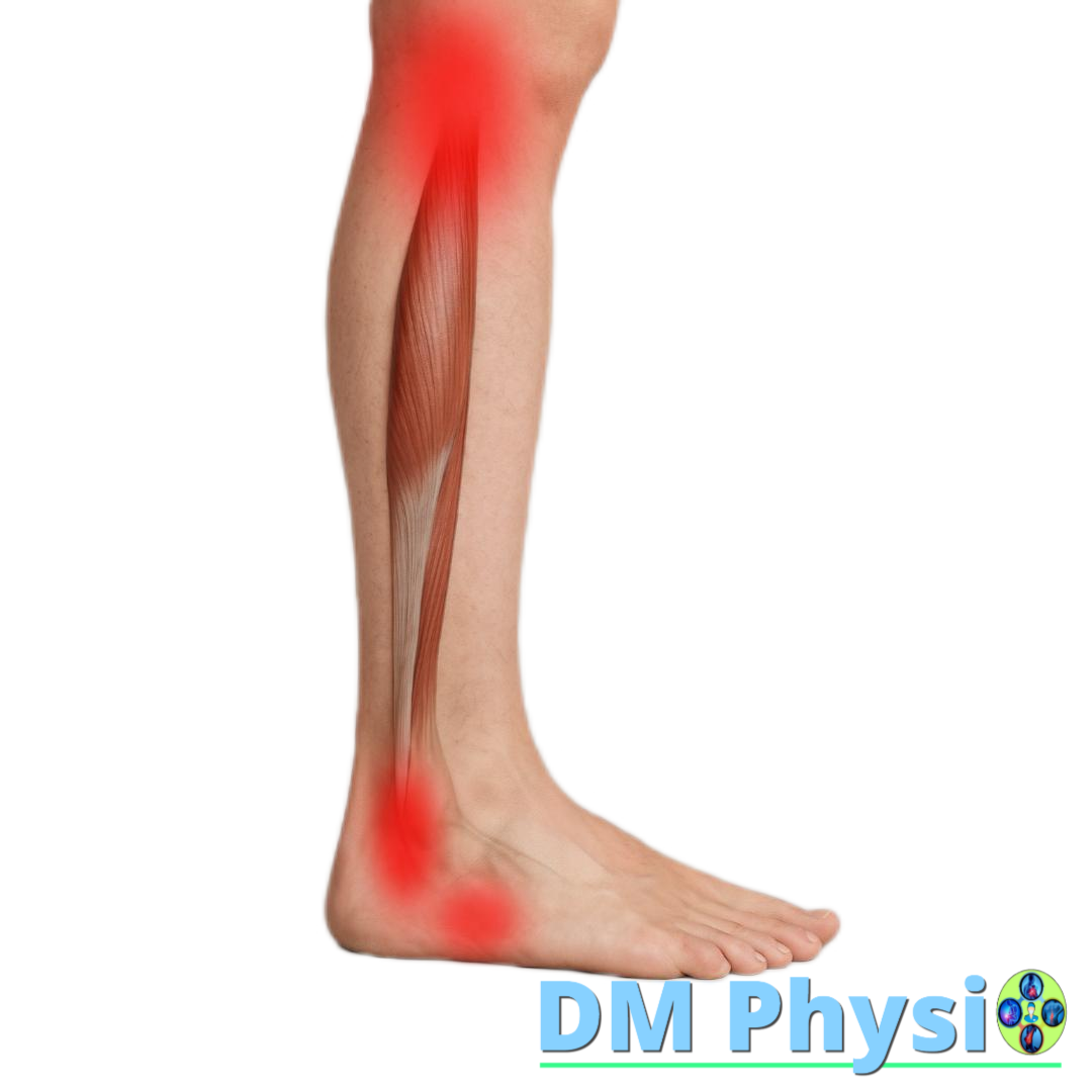
Calf (Triceps surae)
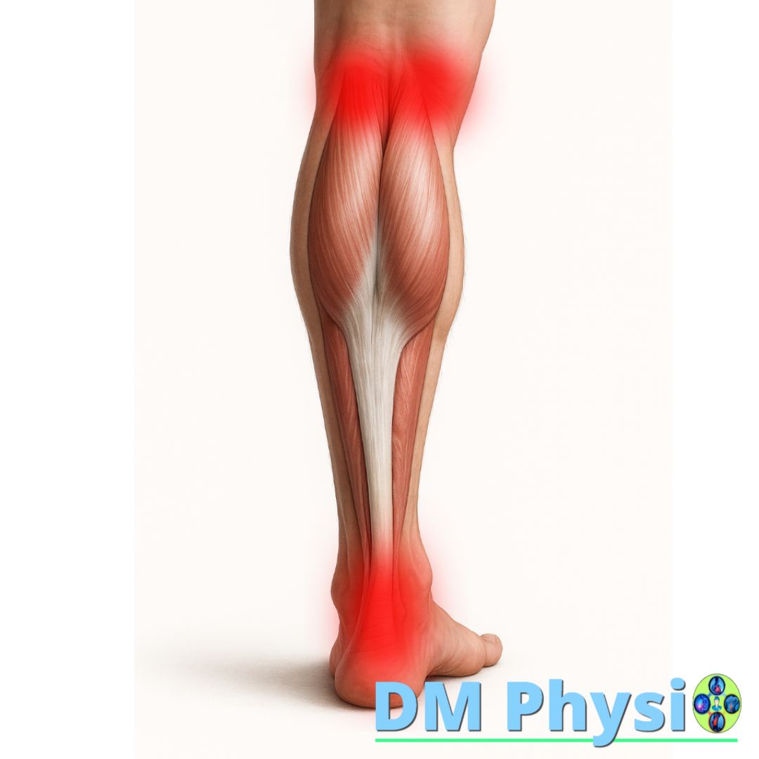
Anatomy and grip
It consists of gastrocnemius and soleus. Both are captured via Achilles tendon for the heel, and the gastrocnemius also passes through the knee.
Nature of the pain
- Calf pain/strain, often to heel and achilles
- Pain behind the knee (medial or lateral)
- Stronger when climbing stairs, walking on tiptoes, and standing for long periods of time
- Frequent morning stiffness and night cramps
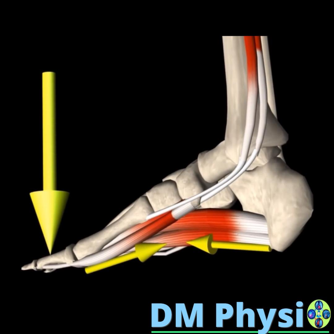
Deep muscles on the inner side of the lower leg
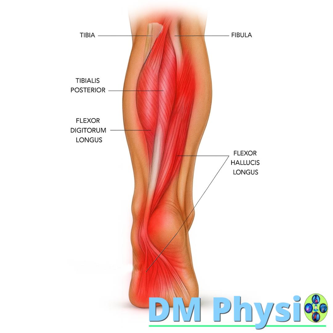
Anatomy and grip
Three muscles whose tendons pass on the inside of the ankle to the arch, fingers and thumb. Tibialis posterior holds the vault; Flexor digitorum longus folds the fingers; Flexor hallucis longus actuates the thrust with the thumb.
Nature of the pain
- Pain from the inside of the ankle or under the arch, sometimes towards the big toe or up the leg
- "Pinching"/clicking with thumb movement (common in FHL)
- Fatigue and heaviness in the arch at the end of the day - a sign of weakness Tibialis posterior
- Aggravation with tight shoes, long standing, or worn soles
Peroneus (Fibularis) - outer lower leg
Anatomy and grip
Tendons pass behind the outer ankle and they keep the ankle from stepping crookedly; often take on more work on rough terrain.
Nature of the pain
- Pain on outer ankle and the outside of the knee, sometimes to the base of the fifth metatarsal
- Clicking/jumping behind outer ankle (tendinous instability)
- Stronger on inclines, lateral movements and downhill running
The muscles in the front of the lower leg (Tibialis anterior, Extensor digitorum longus and Extensor hallucis longus)
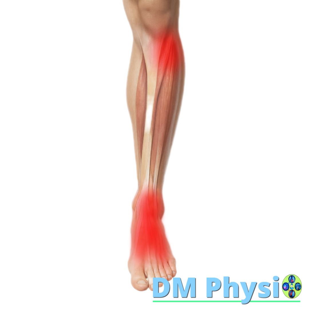
Anatomy and grip
Muscles on the front of the lower leg, which are responsible for lifting the foot and toes up and control smooth heel placement when stepping.
Nature of the pain
- Pain below the knee, around the ankle, or on the top of the foot
- Irritation from tight shoes or stiff tongues
- Often overworked by standing with locked knees for long periods of time
The small extensors of the fingers (Extensor digitorum brevis and Extensor hallucis brevis)
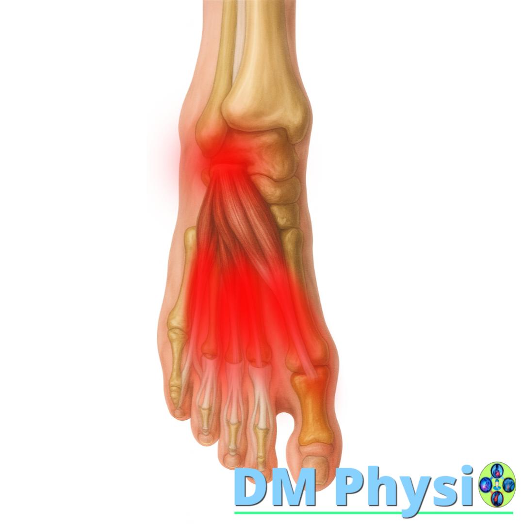
Anatomy and grip
Small muscles on top of foot, near the outside. They help for raising the fingers and thumb up.
Nature of the pain
- Pain on the top of the foot, to the fingers and thumb
- Irritation from tight shoes or stiff tongues, as well as when frequently going down a slope
- Tenderness and tension when trying to straighten the fingers
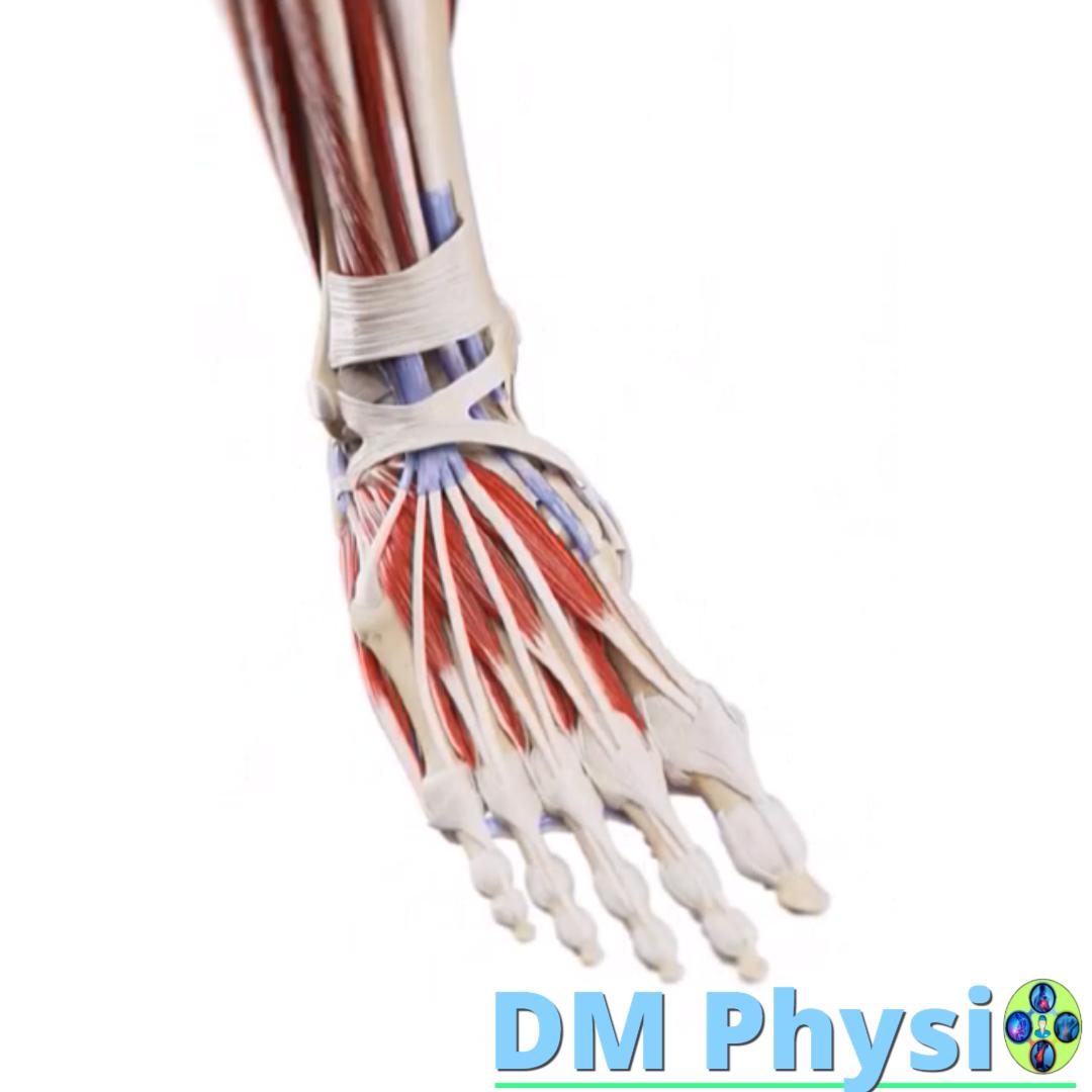
The muscles on the bottom of the foot (Flexor digitorum brevis, Abductor hallucis, Adductor hallucis and Quadratus plantae)
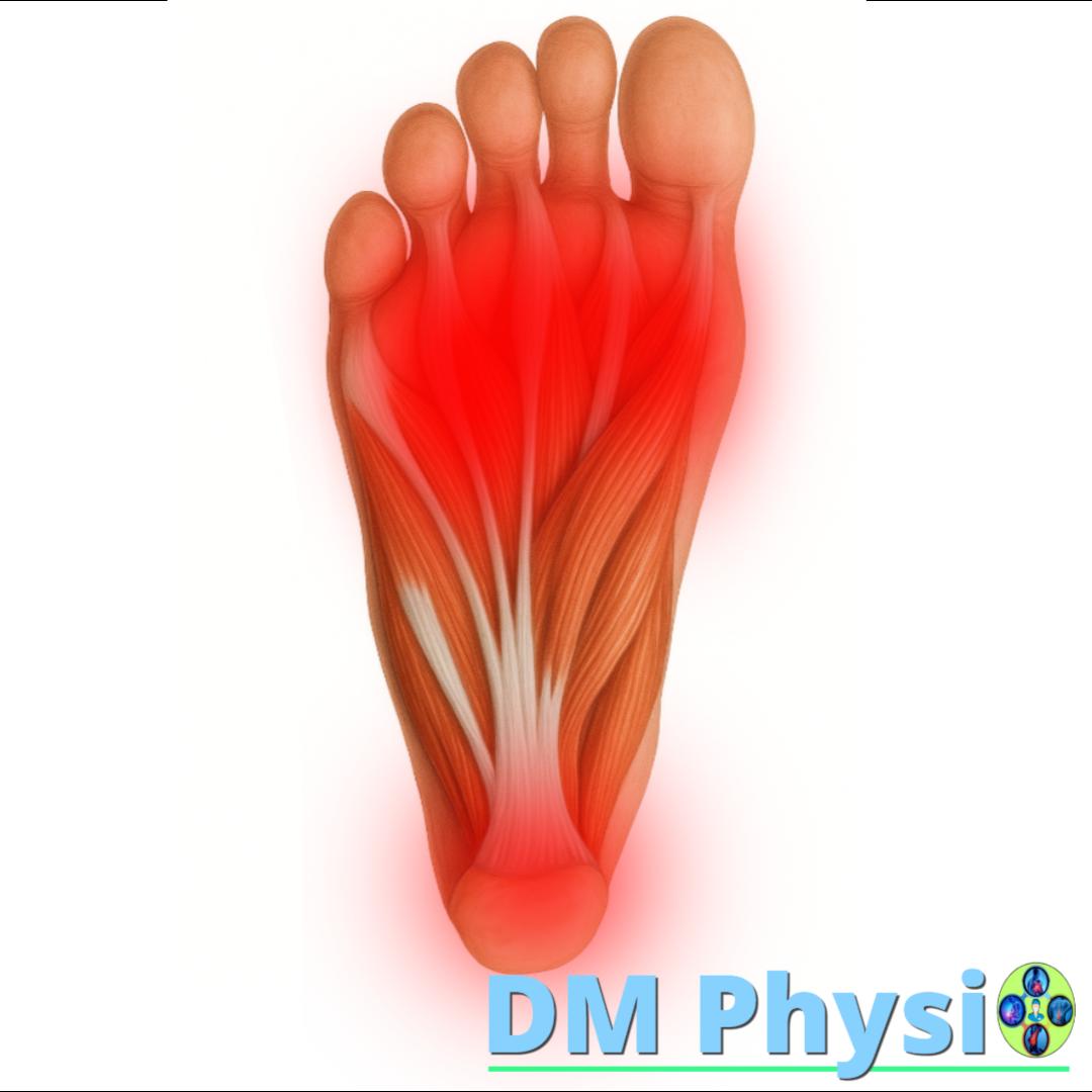
Anatomy and grip
Group plantar muscles on the underside of the foot. They work for finger flexion, arch support and stability when standing and walking.
Nature of the pain
- Arch pain/tension, often confused with plantar fasciitis
- Cramps or stiffness in the fingers, morning tingling, fatigue after standing for a long time
- Stronger symptoms at tight shoes, worn soles or high load
- Their weakness predisposes to deformities such as hallux valgus
How do we reduce pain and improve comfort in the lower leg and foot?
Three steps for better comfort, stability and load tolerance.
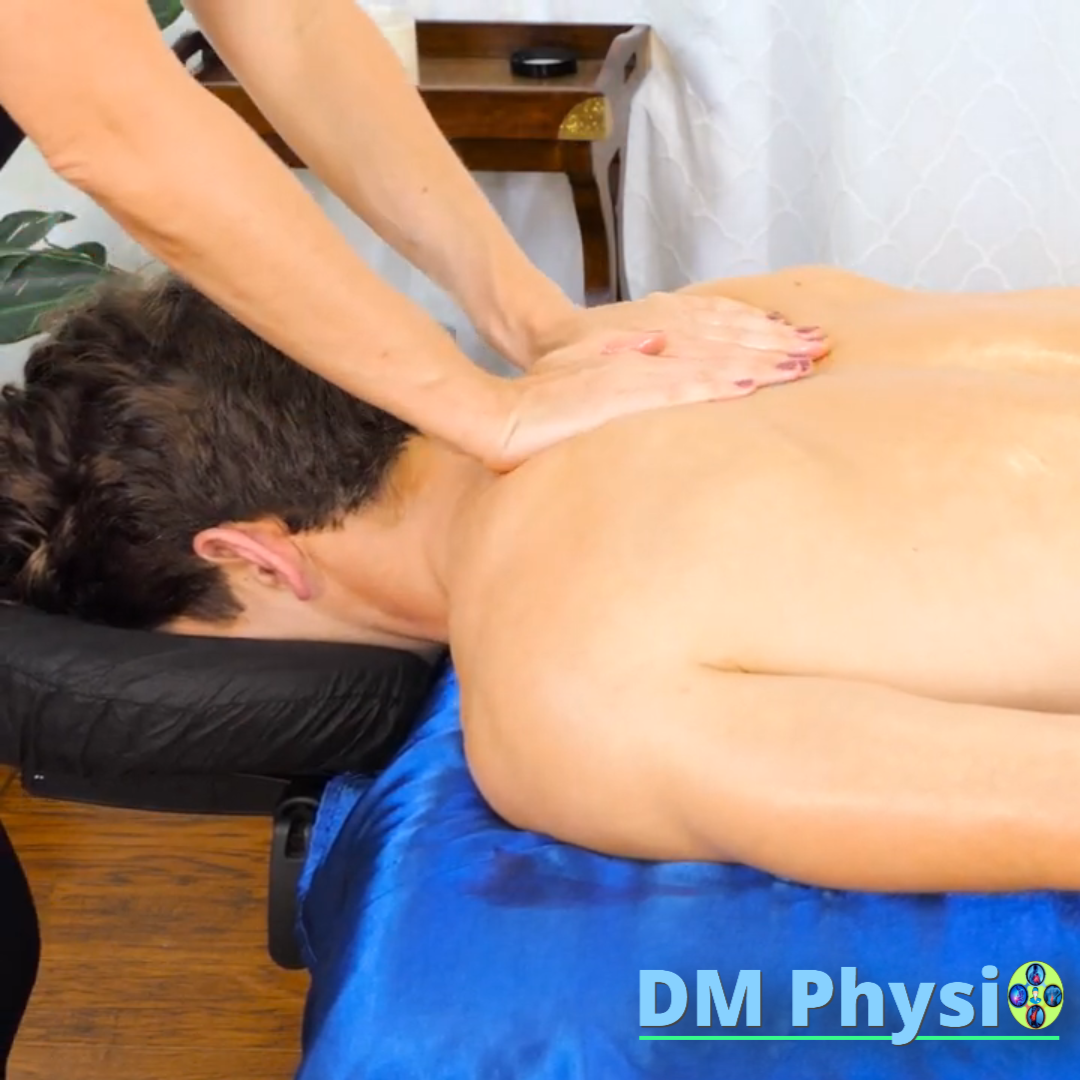
1. Reduce pain
We start with massage and manual techniques, which relax tense muscles and fascia. This can reduce irritation and make movement easier.
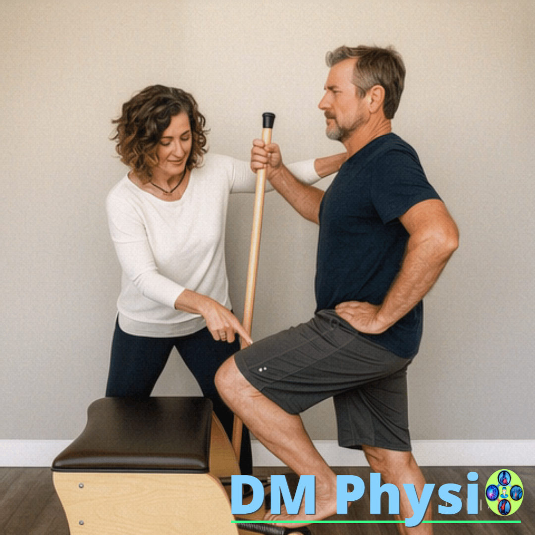
2. Rebalancing
Then we find out muscle imbalances – which muscles are overworked and which are weak. We activate the weakened muscles to restore the stability of the arch and prevent new overload.
3. New movement habits
Finally, we show the difference between wrong and right habits – how to step, what shoes to wear and how to distribute your weight. Thus, the result remains permanent and the pain does not return.